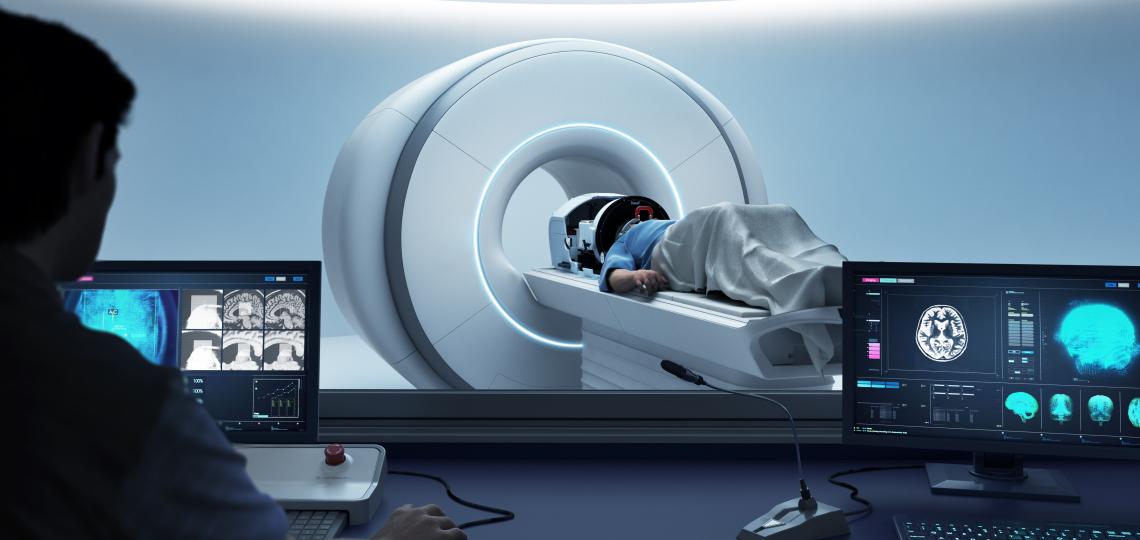
Non-invasive surgery for tremors offers quick relief with low risk
Magnetic resonance-guided focused ultrasound is a treatment option for patients with essential tremor or tremor-dominant Parkinson's disease who have not benefited from medication or are not willing to have deep brain stimulation. The procedure has also recently been approved by the FDA for unilateral treatment of motor fluctuations and dyskinesias in patients with Parkinson’s Disease. The procedure uses focused beams of ultrasonic energy guided by MRI to target areas deep in the brain with no incisions or permanent implants.
Baylor Medicine in partnership with Texas Children's Hospital is one of a few selected centers in the United States with the expertise and technology to provide MR-guided focused ultrasound therapy to patients.
Patients
Patients interested in being evaluated to determine candidacy should call 713-798-4696 or email neurosurgery@bcm.edu. Please review the candidate information below.
Physicians
For physicians interested in learning more about MR-guided focused ultrasound or looking to refer a patient.
Treatment Benefits
- Tremor Improvement: 40-80% improvement in tremor severity (many with immediate improvement) stably maintained beyond five years.
- Incisionless: Focused ultrasound technology allows sound waves to pass safely through the skull without incisions. No implants and no radiation required.
- Quick Recovery: With no surgical cuts, there is minimal to no risk of infection. The treatment is often performed on an outpatient basis with local anesthesia, and you can expect to resume normal activities within days.
- Safe and Effective: FDA-approved treatment provides real-time thermal feedback to continuously monitor patient safety and temperature at the target site with minimal side effects.
How it Works
The concept of MR-guided focused ultrasound is much like using a magnifying lens to focus the sun's energy on a leaf. If you put your hand under the magnifying lens, you feel no heat, but the concentrated energy is strong enough to burn the leaf at the focal point. For the treatment of tremor, focused ultrasound therapy is applied on deep brain structures, creating a tiny ablation or lesion (like the pinpoint burn on the leaf). This small lesion created by focusing ultrasound in a specific brain region interrupts the transmission of abnormal brain activity responsible for tremor, leading to tremor reduction and improvements in quality of life. The FDA has approved the use of magnetic resonance-guided focused ultrasound to treat patients with essential tremor or and tremor-dominant Parkinson's disease. More recently, the procedure has been approved for unilateral treatment of motor fluctuations and dyskinesias in patients with Parkinson’s Disease.
Patient Candidacy
It's extremely important to discuss all medical conditions with your physician to evaluate your suitability for the procedure properly. All patients considering focused ultrasound treatment must undergo a screening process. Generally, the steps in the focused ultrasound screening process include:
- Discussion and process initiation with your neurologist.
- Motor evaluation, videotaping of your movement examination, and overall health assessment.
- Your may need to undergo a neuropsychological evaluation to determine various motor and cognitive abilities.
- Computed tomography (CT) scan to measure skull thickness for focused ultrasound calculations.
- Discussion of your focused ultrasound candidacy in a multi-disciplinary conference consisting of a team of neurologists, a nurse practitioner, neuropsychologists, and neurosurgeons.
- Clinic visit with the neurosurgeon who will perform your focused ultrasound treatment to discuss the procedure.
Treatment Day
Preparation
On the treatment day, we begin by giving you a close haircut. This head shave is necessary for the ultrasound waves to be appropriately transmitted through the skin and skull into the target region in the brain. Local anesthesia will be applied to numb areas of your scalp, and a frame will be secured to your head so that your head doesn’t move during the treatment. We then attach a helmet to the frame. Cool water will circulate in the helmet around the top of your head to minimize potential heating near the scalp.
Your heart rate, blood pressure, and blood oxygen levels will be monitored throughout the procedure. You may be given additional medication to keep you comfortable.
You will also be given a “stop sonication” button to indicate to the physician that you want to stop the treatment for any reason.
Planning and Procedure
You will then lie down in the MRI scanner equipped with the focused ultrasound machine. A series of MRI images will be taken for planning the treatment according to your specific anatomy. The treating neurosurgeon will first apply light doses of ultrasound energy and real-time images of the ultrasound delivery will be taken. After each application of energy, called a sonication, you will be asked to perform specific tasks to evaluate your tremor improvement. Tasks may include drawing spirals on a board or performing tasks with your hands. The team will continue to fine-tune the therapy and identify any side effects. The treating neurosurgeon will then apply higher energy to create the permanent lesion. It is common to experience a noticeable reduction in tremor during the procedure itself. At the end of the procedure, a final MRI scan will be done to assess the treatment. The procedure will last approximately between 3-4 hours.
After Treatment
After the procedure, you'll be transferred to a recovery room for a short monitoring period. The frame will be removed. The physician will let you know when you can go home. Within days you should be able to return to normal activities.


