

This is the main microscope room containing a Leica confocal, a Zeiss Axiophot, a Nikon SMZ1500 dissecting microscope.

The Zeiss Axiophot microscope is capable for bright field and epifluorescent imaging with both black and white, and color CCD cameras.
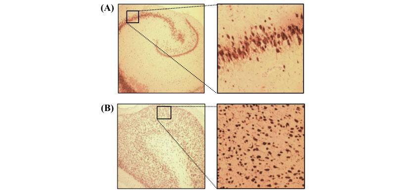
Bright field imaging from the Zeiss microscope of brain sections stained with a neuronal marker to test for differences in brain structure among pre-mature baby pigs given different formulations of TPN. These images were captured using a SPOT-SliderRT CCD camera.

The Leica SME Laser Scanning Confocal Microscope images 4 channels with excitation wavelengths of 405, 488, 563, and 633 nm. Channels are sequentially imaged using a tunable emission filter. The system is capable of automated tiling for multiple fields or image stacks as well as collection of time series data.
This scope has a fully motorized stage, which facilitates both the image tiling functions as well as the automated collection of z-stacks. Image brightness, background intensity, and focal plane, are all controlled through a digital console behind the keyboard.
This scope is integrated into an image analysis workstation that consists of a DELL Optiplex GX620 processor with a Radeon X600SE video card, Adobe Photoshop image processing software and ImagePro 10.0 image analysis software. The image analysis software consists of a basic analysis module with plug-in modules for image deconvolution (Sharp Stack Plus Ver 5.0), and 3 dimensional reconstruction (3D Constructor).

The Nikon SMZ 1500 dissecting microscope is capable of imaging bright-field and fluorescent specimens from 70x to 150x. The scope also has flexible top illumination to facilitate microdissections.

Meyer Slide scanner quickly collects images of stained tissues sections allowing a global view of tissue architecture.

Olympus Fluoview confocal microscopy system consisting of an IX70 microscope, four lasers (blue diode, argon, green HeNe, and red HeNe) a laser combiner and FV300 confocal scanner, a bright-field detector, a Spot RT color CCD camera, a computer work station, and a Kodak 8600 dye-sublimation printer


ZEISS Lightsheet 7 (Room 8058)
The ZEISS Lightsheet 7 is optimized for rapid, gentle 3D imaging of large, optically cleared tissue specimens (compatible with SmartBatch+ clearing) as well as live organisms. Its specialized optics accommodate refractive indices ranging from 1.33 to 1.58. The system utilizes dual-side sheet illumination with decoupled illumination and detection beam paths to minimize phototoxicity and photobleaching during extended live acquisitions. Multiview sample rotation facilitates full angular coverage and 3D volumetric fusion across four detection channels (DAPI, GFP, RFP, and Far Red).

Oxford Instruments Andor BC43 Benchtop Confocal Microscope (Room 8058)
The Andor BC43 is a benchtop multimodal imaging system combining high-speed spinning-disk confocal microscopy (up to 10× faster than conventional point-scanning confocal), widefield epifluorescence, and differential phase contrast (DPC)/brightfield imaging. Equipped with 10X to 60X objectives and four laser lines (405, 488, 561, and 638 nm), it supports deep tissue imaging in organoids, embryos, and model organisms with automated tile stitching. The system integrates GPU-accelerated deconvolution and 3D visualization, offering high-sensitivity, low-phototoxicity acquisition ideal for dynamic live-cell and fixed specimen studies.

ZEISS Axio Scan.Z1 Slide Scanner (Room 8058)
The ZEISS Axio Scan.Z1 is an automated digital slide scanner engineered for high-throughput brightfield and multichannel fluorescence imaging. Capable of loading up to 12 standard slides per automated run, it is equipped with a high-efficiency LED light engine and 5X, 10X, and 20X objectives. The system provides seamless whole-slide digitization for global tissue histology overviews and quantitative fluorescence mapping of large histological sections.

Leica DM5500B Upright Widefield Microscope (Room 10059)
The Leica DM5500B is an automated research upright microscope supporting brightfield, darkfield, phase contrast, polarization, and Differential Interference Contrast (DIC) imaging modalities. It features a motorized 6-objective nosepiece (2.5X, 5X, 10X, 20X, 40X, and 63X) alongside multichannel widefield epifluorescence configured for DAPI, GFP, Cy3, and Cy5 filter sets, providing versatile high-resolution imaging for stained tissue sections and cell culture preparations.
Questions
Questions concerning suitability of the system for particular applications can be directed to Dr. Gregory Guthrie associate professor Pediatrics and Molecular and Cellular Biology.