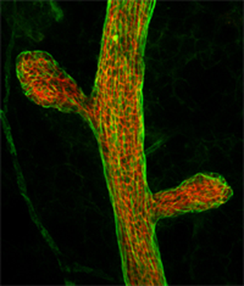
The Confocal Microscopy and Image Analysis Core provides instrumentation and expertise to support the acquisition, processing and analysis of both fluorescence and bright-field micrographs captured from fixed tissue sections, whole mounts, or from either fixed or live cells.
Digital micrographs can be captured from any one of four different microscopes including an Olympus FV300 Laser Scanning Confocal Microscope capable of collecting 3-dimensional or time series data on specimens containing up to four separate fluorochromes.
An image processing and analysis workstation provides tools to process and collect data from digital micrographs. The capabilities of this system include deconvolution, co-localization, 3-dimensional reconstruction, pixel-intensity measurements, and morphometry. Analysis can be conducted using both manual and automated approaches.
Custom cryosectioning, staining, image acquisition and analysis are also available.
Core Operations
The Confocal Microscopy and Image Analysis Core facility will be available to both USDA/ARS Children's Nutrition Research Center personnel as well as college-wide within Baylor as a fee-for-service facility.
New users, or users unfamiliar with the use of the system will be required to attend an initial two hour training session to familiarize them with the system. If the user is a junior investigator, post-doctoral fellow, graduate student or technician, the user’s principle investigator is also required to attend this initial training session.
After completing the training session, new users will be required to complete a minimum of 10 hours of supervised use before independent confocal use can begin.
All users of the microscope will be required to fill out a log sheet that will be kept near the microscope. Failure to complete the log sheet will result in loss of privileges.
All scheduling of instruments will be done through the Fluoview calendar on Outlook webmail. You must schedule your use no later than noon on the day before you intend to use the instrument. For weekend and Monday use this means no later than noon on Friday. When scheduling, you will need to provide your email address, your PI, and the instrument you intend to use. Access the calendar as described below.
Scheduled Users
A printout of scheduled users will be posted each day at noon for the following day. Access the Outlook webmail through the Baylor College of Medicine intranet. In Outlook:
- Click ‘Folder List’ which is at the bottom left of the Outlook screen.
- Click the + before ‘Public Folders’ then click the + before ‘All Public Folders’.
- Scroll down to ‘BCM Pediatrics’ then inside to ‘Nutrition’. Click ‘Fluoview Schedule’.
- To add the calendar to your favorites, right-click the calendar and then click ‘Add to Favorites’.
- Click ‘Calendar’ (beside ‘Folder List’ at the bottom left of the Outlook screen).
- You should see this new calendar listed under ‘Other Calendars’.
Activity and Billing
Principle investigators will receive periodic statements of activity along with a bill for this activity.
Scheduling and Questions
Scheduling training or questions concerning suitability of the system for particular applications can be directed to Dr. Yongjie Yang.


