About the Program
Next opening: 2028-2029
Number of positions: 1
Length: 1 year
This ACGME-accredited fellowship provides robust training in disorders of the luminal GI tract, pancreas and hepatic pathology (medical liver including liver transplantation), preparing trainees for a career in an academic or private practice setting. Five GI pathologists provide clinical service, contribute to the research program of the GI section, and support the GI Pathology training program across four distinct hospital and outpatient settings.
The GI pathology fellow will get exposure to a comprehensive spectrum of gastrointestinal surgical specimens, ranging from small biopsies to complex procedures, as well as hepatobiliary specimens. Training also includes reviewing consult digital slides from community practices in Texas. The fellow will have graduated responsibility in performing operating room consultations (frozen sections and gross examination). The fellow is expected to closely interact with surgeons/clinicians and pathology colleagues from community practice, actively participate in tumor boards, IBD/liver conferences, participate in research projects and will have the opportunity to attend and contribute to numerous didactic and clinical conferences.
Electives in clinical gastroenterology are available.
Rotations
Fellows will rotate 9 months at Baylor St. Luke’s Hospital, 1 month at Ben Taub General Hospital, 1 month at Texas Children’s Hospital, and 1 month at MD Anderson Cancer Center.
The fellow has the option to rotate for 2 weeks in the Gastroenterology Department, to observe upper and lower endoscopies and EMR/ESD procedures.
Stipends and Benefits
Visit the Graduate School of Biomedical Sciences page on stipends and benefits for fellows.
Admissions
Candidates must be board certified or eligible for certification in anatomic pathology or AP/CP.
All application materials, including recommendation letters, must be submitted online, and include the following:
- Completed online House Staff Application
- Current Curriculum Vitae
- Personal Statement
- Transcript from Medical School(s)
- Three Recommendation Letters
Applications are accepted from mid-April to July 31 for positions beginning July 1 two years later. Interviews are conducted starting in July or August and are completed by the end of August of the application year.
Case of the Month
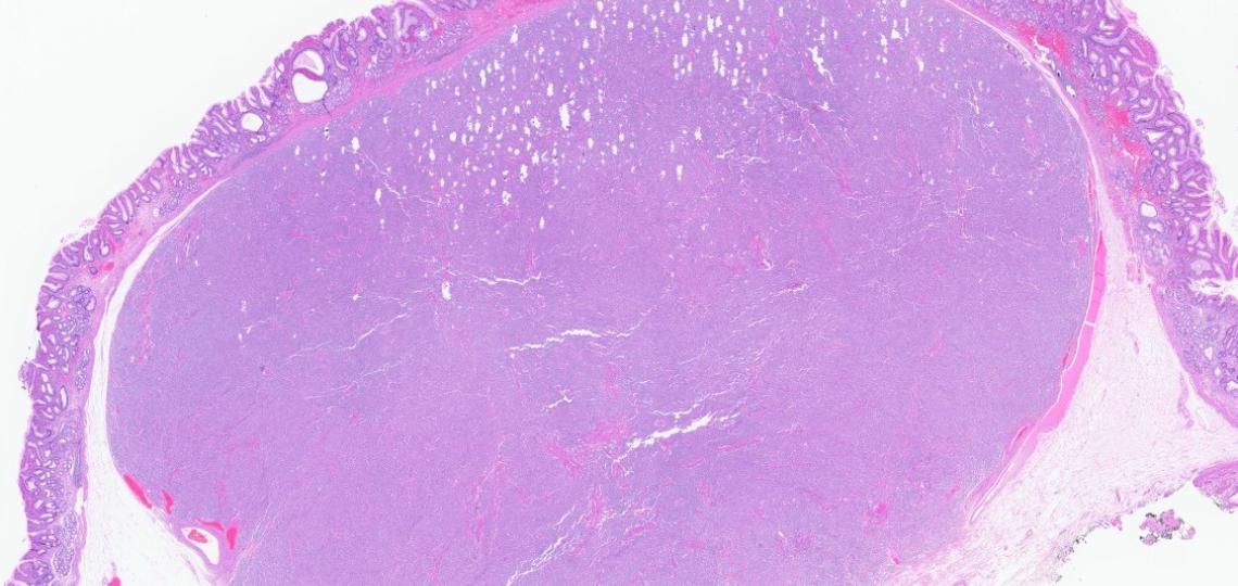
Question 1: Which of the following histologic patterns is associated with a higher grade?
- A: Cribriform
- B: Tubular
- C: Solid
This tumor is not graded based on histopathologic features
Answer: C (Solid).
Explanation for Q1:
The diagnosis is Adenoid cystic carcinoma
Adenoid cystic carcinoma has an incidence of 1–2 cases per 100 000 person-years, making up 20% of all malignant salivary gland tumors. Patients present with neural symptoms or a slow-growing mass, most commonly in the parotid, submandibular, and minor glands of the palate. Lymph node metastasis is uncommon. However, a prolonged clinical course with local recurrences and distant metastases to the lungs, bone, liver and brain are commonly seen. Overall survival rate at 10 years is 50%, with a median survival time of 36 months after diagnosis.
Two main cell types are found in this tumor: ductal cells, with an eosinophilic cytoplasm and uniform round nuclei, and myoepithelial cells with a clear cytoplasm and hyperchromatic angular nuclei. In ACC, ductal cells are strongly positive for CK7 and CD117. Myoepithelial cells are positive for p63, p40, calponin, and α-SMA. Perineural invasions are commonly found.
Histologically, three architectural patterns are recognized, tubular, cribriform, and solid, and a combination of all three is common. The tubular pattern consists of ducts and tubules lined with luminal ductal and abluminal myoepithelial cells. The cribriform pattern is characterized by nests of tumor cells with microcystic-like spaces filled with hyaline or basophilic mucoid material. The solid pattern is characterized by tumor sheets of basaloid without a specific architecture.
Recent studies have shown that the presence of any solid tumor component indicates a higher grade. The clinical course is more aggressive when > 30% of the solid component is found. Other factors that affect survival include patient age, tumor site, TNM stage, surgical margins, and NOTCH1 mutation status.
Q2: Which genetic alterations are most commonly found in adenoid cystic carcinoma?
- A: CRTC1–MAML2 fusion
- B: MYB::NFIB fusion
- C: MYBL::NFIB fusion
- D: PLAG1 rearrangements
Answer: B and C (MYB::NFIB and MYBL1::NFIB fusion).
Explanation for Q2:
Salivary ACC commonly harbors a t (6;9) chromosomal translocation, resulting in fusions involving the MYB or MYBL1 and NFIB genes (>90% of cases).
In our case, a universal solid tumor NGS fusion panel was performed, and no fusion was detected. This does not exclude the diagnosis of ACC. Since the mass was in the periesophageal space, centered in the posterior tracheal wall and the anterior wall of the esophagus, it is uncertain where it originates from. It could be arising from the trachea and extending towards the esophagus, or it could be arising from the esophageal submucosa.
Primary tracheobronchial adenoid cystic carcinoma is rare (<1% of all lung tumors) but still more common than primary esophageal ACC (0.1%). Previous studies have reported an MYB::NFIB fusion in 41-50% of tracheobronchial ACCs. Another case series (n=7) has shown the presence of fusions involving MYB or MYBL1 and NFIB in 100% of cases 4. Given the higher incidence of ACC in the respiratory tract and the absence of detectable fusion, determining the exact origin of the tumor in this biopsy specimen remains challenging. Additionally, since the patient has no history of other tumors, metastasis is unlikely.
References
van Weert S, van der Waal I, Witte BI, Leemans CR, Bloemena E. Histopathological grading of adenoid cystic carcinoma of the head and neck: analysis of currently used grading systems and proposal for a simplified grading scheme. Oral Oncol. 2015;51(1):71-76. doi:10.1016/j.oraloncology.2014.10.007
Morita N, Murase T, Ueda K, et al. Pathological evaluation of tumor grade for salivary adenoid cystic carcinoma: A proposal of an objective grading system. Cancer Sci. 2021;112(3):1184-1195. doi:10.1111/cas.14790
Brayer KJ, Frerich CA, Kang H, Ness SA. Recurrent Fusions in MYB and MYBL1 Define a Common, Transcription Factor-Driven Oncogenic Pathway in Salivary Gland Adenoid Cystic Carcinoma. Cancer Discov. 2016;6(2):176-187. doi:10.1158/2159-8290.CD-15-0859
Pei J, Flieder DB, Patchefsky A, et al. Detecting MYB and MYBL1 fusion genes in tracheobronchial adenoid cystic carcinoma by targeted RNA-sequencing. Mod Pathol. 2019;32(10):1416-1420. doi:10.1038/s41379-019-0277-x
Yoshikawa K, Kinoshita A, Hirose Y, et al. Endoscopic submucosal dissection in a patient with esophageal adenoid cystic carcinoma. World J Gastroenterol. 2017;23(45):8097-8103. doi:10.3748/wjg.v23.i45.8097
Inagaki H, Faquin WC, Stenman G, Urano M. Adenoid cystic carcinoma. In: WHO Classification of Tumours Chapter 4: Salivary gland tumours. 5th ed. International Agency for Research on Cancer (IARC)
1. A 55-year-old woman undergoes an appendectomy for a suspected appendiceal mass. Histopathologic examination reveals an infiltrative neoplasm composed of clusters and small nests of goblet cells with areas of extracellular mucin production. Immunohistochemistry shows focal positivity for synaptophysin and chromogranin in the goblet cells.
Which of the following is the most appropriate interpretation of the neuroendocrine marker expression? (Select one answer)
- A. Indicates a high-grade neuroendocrine carcinoma (NEC), warranting chemotherapy
- B. Suggests a mixed adenoneuroendocrine carcinoma (MANEC) requiring dual component quantification
- C. Is a known feature of goblet cell adenocarcinoma
- D. Confirms the diagnosis of well-differentiated neuroendocrine tumor (NET) of the appendix
- E. Reflects non-specific staining and should be disregarded in diagnosis
Answer: C(Correct). Focal and weak positivity for synaptophysin and chromogranin is a known feature of goblet cell adenocarcinoma
Explanation: Goblet cell adenocarcinoma (previously called goblet cell carcinoid) is a unique neoplasm of the appendix that exhibits features of both adenocarcinoma and neuroendocrine differentiation. However, the expression of neuroendocrine markers such as synaptophysin and chromogranin can be focal or weak and does not imply true neuroendocrine differentiation or behavior. Moreover, even when INSM1 (a sensitive neuroendocrine marker) is positive, staining is typically limited to a small minority of tumor cells (mean ~8%), underscoring that expression is focal and not diagnostic of a neuroendocrine neoplasm. Unlike classical neuroendocrine tumors, GCAs behave more aggressively and are treated like adenocarcinomas. Misinterpreting this marker positivity could lead to inappropriate treatment choices.
2.Which of the following features help differentiate goblet cell adenocarcinoma (GCA) from signet-ring cell adenocarcinoma (SRCA)?
(Select one answer)
- A. Presence of a low-grade goblet cell component
- B. Widespread (>50%) discohesive signet-ring cells
- C. High mitotic activity and necrosis
- D. Cribriform growth pattern with extracellular mucin
Answers: A (Correct). The presence of a low-grade goblet cell component. Goblet cell adenocarcinoma is defined by the presence of a recognizable low-grade goblet cell adenocarcinoma component.
Explanation: Signet-ring cell adenocarcinoma shows widespread (> 50%) discohesive and disorganized growth of signet-ring cells with high-grade cytological features and lacks a low-grade goblet cell component.
While high-grade areas of GCA can show numerous atypical mitotic figures and necrosis, these features are not exclusive to SRCA and can be seen in both high-grade GCA and SRCA. Therefore, they do not help differentiate between the two.
The cribriform pattern and extracellular mucin are features that can be present in high-grade GCA, but they are not specific enough to distinguish it from SRCA. SRCA is defined more by its discohesive signet-ring cells and lack of low-grade GCA component, not by architectural patterns like cribriform growth. Extracellular mucin is often present and sometimes abundant in SRCA.
Goblet cell adenocarcinoma diagnostic criteria and grading
Low grade features
Required: Classic round tubular growth pattern composed of predominantly of goblet-like mucinous cells and a fewer number of Paneth-like cells and endocrine-like cells
Common : Extracellular mucin pools containing round tubules or cohesive clusters, including ruptured tubules
Uncommon: Tubules with non mucinous glands, including oncocytic tubules
High grade features: 5 patterns
Signet ring-like/goblet-like cells diffusely infiltrating predominantly as single cells or abortive tubules.
Signet ring-like/goblet-like cells with fusion of clusters to form large complex anastomosing structures or very large aggregates.
Infiltrative single-file or anastomosing cords of tumor cells with high-grade nuclei. Intracytoplasmic mucin may be only very focally present.
Gland-forming adenocarcinoma, often with a microglandular growth pattern of tubules with high-grade nuclei. Intestinal-type adenocarcinoma is less common.
Solid sheet-like growth of tumor cells with high-grade nuclei and minimal intracytoplasmic mucin.
Grading: 3 tier grading system based on low grade versus high grade patterns
Reference:
WHO Classification of Tumours Editorial Board.
Digestive System Tumours (5th edition). IARC, 2019.
Appendix tumors are reclassified; GCA is considered distinct from NETs and NECs.
Roy P, Chetty R. Goblet cell carcinoid tumors of the appendix: An overview. World J Gastrointest Oncol. 2010 Jun 15;2(6):251-8. doi: 10.4251/wjgo.v2.i6.251. PMID: 21160637; PMCID: PMC2998842.
Lin DL, Wang LL, Zhao P, Ran WW, Wang W, Zhang LX, Han M, Bao H, Liu K, Wu X, Shao Y, Xing XM. Gastrointestinal Goblet Cell Adenocarcinomas Harbor Distinctive Clinicopathological, Immune, and Genomic Landscape. Front Oncol. 2021 Nov 5;11:758643. doi: 10.3389/fonc.2021.758643. PMID: 34804955; PMCID: PMC8603204
Question 1. Which statement is true about gastrointestinal-originated spindle cell neoplasms? (More than one statement can be true)
A. Significant CD117 and DOG1 positivity in gastrointestinal spindle cell tumors rules out the diagnosis of leiomyoma.
B. Interstitial cells of Cajal can only exhibit focal DOG1 positivity.
C. The presence of cytoplasmic dendritic extensions is in favor of ICCs.
D. Entrapped ICC cells in leiomyomas can be only seen in deep leiomyomas.
E. The presence of ICCs in esophageal leiomyoma causes more significant clinical implications compared to gastric leiomyoma.
F. Mast cells usually mimic the same histological findings that can be observed in ICCs entrapped in leiomyoma.
Answer: C and E are true statements.
Explanation:
Interstitial cells of Cajal (ICCs) are specialized mesenchymal cells that serve as pacemakers for gastrointestinal motility and are thought to play a key role in neuromuscular transmission. Although all subtypes of ICCs share fundamental ultrastructural characteristics, their morphology can vary depending on species and anatomical location, ranging from fibroblast-like to smooth muscle cell-like appearances.
Distinguishing gastrointestinal stromal tumors (GISTs) from leiomyomas can pose a significant histopathological challenge, especially in fine-needle aspiration or core biopsy specimens. This distinction is particularly crucial in the esophagus, where therapeutic approaches differ markedly. Leiomyomas, even when large, are often amenable to conservative management such as enucleation, thereby avoiding more invasive procedures like esophagogastrectomy—an operation associated with considerable morbidity and a small but real mortality risk. In contrast, esophageal GISTs, the majority of which exhibit malignant potential, typically necessitate more radical surgical intervention, such as esophagectomy. In the stomach, however, the therapeutic distinction is less critical, as both GISTs and leiomyomas are generally treated with conservative resection.
The diagnostic complexity is further compounded by the observation that some gastrointestinal leiomyomas contain cells that resemble interstitial cells of Cajal. These ICC-like cells are morphologically similar to native ICCs, characterized by cytoplasmic dendritic processes and strong immunoreactivity for DOG1 and KIT. They are most identified in deep-seated leiomyomas, reflecting the anatomical distribution of ICCs within the muscularis propria. Nonetheless, rare reports have documented their presence in more superficial lesions as well, supporting the hypothesis that smooth muscle cells and ICCs may originate from a common progenitor, possibly reflecting a spectrum of stromal tumors with mixed differentiation.
Mast cells—another population of KIT-positive cells—may also be present within smooth muscle tumors. However, they can be distinguished from ICCs by their oval shape, dense chromatin, and limited numbers. In contrast, ICCs are more diffusely distributed and possess elongated, branching processes.
Careful histological assessment of KIT-positive cells, with attention to their morphology, can aid in differentiating ICCs from mast cells. Immunohistochemical profiling also plays a critical role. Extensive and diffuse positivity for smooth muscle markers such as desmin and smooth muscle actin (SMA) supports a diagnosis of leiomyoma, even when patchy areas of reduced expression are present. This is reinforced by the fact that desmin positivity is rare in GISTs—reported in only about 5% of gastric and up to 15% of esophageal cases.
References:
Deshpande A, Nelson D, Corless CL, Deshpande V, O'Brien MJ. Leiomyoma of the gastrointestinal tract with interstitial cells of Cajal: a mimic of gastrointestinal stromal tumor. Am J Surg Pathol. 2014 Jan;38(1):72-7. doi: 10.1097/PAS.0b013e3182a0d134. PMID: 24145645.
Janevska V, Qerimi A, Basheska N, Stojkova E, Janevski V, Jovanovic R, Zhivadinovik J, Spasevska L. Superficial leiomyomas of the gastrointestinal tract with interstitial cells of Cajal. Int J Clin Exp Pathol. 2015 Dec 1;8(12):15977-84. PMID: 26884872; PMCID: PMC4730085.
Q: A 57-year-old female with a resected pancreatic mass. All of the below sentences are true about this entity except?
A. On imaging, it may present as cystic lesions in the pancreatic groove.
B. It is often misdiagnosed clinically as cancer arising either in the duodenum, distal CBD, or pancreas.
C. Commonly associated with ductal adenocarcinoma.
D. More frequent in males with history of alcohol abuse and smoking.
Answer: C. Commonly associated with ductal adenocarcinoma
Explanation: Adenocarcinoma is very rarely associated with Groove Pancreatitis (GP). In this case, a 1mm focus of invasive adenocarcinoma was identified next to the ampulla. The ampulla also showed focal low and high-grade dysplasia. Please see images (circled).
- Groove pancreatitis (GP) is a unique form of chronic pancreatitis affecting the pancreatoduodenal groove, a potential space between the head of the pancreas, duodenum, and common bile duct (CBD).
- On imaging, it may present as either mass-like lesions or cystic lesions in the pancreatic groove, mimicking malignancy. Discerning between GP and pancreatic cancer is often challenging but clinically very important.
- Histology shows marked fibrosis of the pancreaticoduodenal groove, dilated and ruptured ducts containing proteinaceous material, acute and chronic inflammation, giant cell reaction, and Brunner gland hyperplasia.
- GP is rare and often misdiagnosed clinically as cancer arising either in the duodenum, distal CBD, or pancreas. GP is more frequent in males and is associated with alcohol abuse, smoking, and anatomical or functional obstruction of the minor papilla.
- Infrequently, adenocarcinoma may be present in the duodenal groove, making differential diagnosis with GP difficult. GP may be treated conservatively, but surgery is indicated when malignancy is suspected and/or symptoms get worse.
- Identifying typical features of GP in a Whipple specimen should not preclude the pathologist from a thorough examination for adenocarcinoma.
References:
- Kristjan Ukegjini, Thomas Steffen et al. Systematic review on groove pancreatitis: management of a rare disease, BJS Open, Volume 7, Issue 5, 2023, October;7(5):1-8.
- E. Fagundo, E. Weisberg, E. Fishman, Pancreatic cancer in patient with groove pancreatitis: Potential pitfalls in diagnosis, Radiology Case Reports,2022, December;17 (12):4632-4635.
- T. Tarvainen, T. Nykänen et al. Diagnosis, natural course, and treatment outcomes of groove pancreatitis, Hepato-pancreato-biliary journal, 2021; August;23(8):1244-1252.
- Koji Yamaguchi, Masao Tanaka, Groove pancreatitis masquerading as pancreatic carcinoma: The American Journal of Surgery, 1992, March; 163(3): 312-316.
- Raman SP, Salaria SN, Hruban RH, Fishman EK. Groove pancreatitis: spectrum of imaging findings and radiology-pathology correlation. AJR Am J Roentgenol. 2013 Jul;201(1):W29-39. doi: 10.2214/AJR.12.9956. PMID: 23789694; PMCID: PMC4005339.
Q1: Which of the following morphological features most strongly suggest a metastatic tumor rather than a primary tumor origin?
A) Adenocarcinoma without any overlying precursor lesion
B) Extensive lymphovascular invasion in lamina propria or superficial
submucosa
C) Endoscopic and radiological correlation
D) All of the above
Answer: D. All of the above.
Explanation for Q1:
The biopsy of the rectal mass demonstrated infiltrating irregular glands within the submucosa. Importantly, the overlying epithelium was unremarkable, showing no evidence of dysplasia (refer to the PowerPoint image).
While metastatic adenocarcinoma to the rectum may occasionally involve the surface epithelium through cancerization, this case lacked any precursor lesions such as tubular adenoma or high-grade dysplastic changes.
The presence of extensive lymphovascular invasion in the lamina propria and superficial submucosa supports a metastatic origin rather than a primary rectal neoplasm. Correlation with endoscopic findings and imaging studies is crucial before finalizing the diagnosis.
Histologically, the tumor exhibits glands lined by cuboidal cells with a low nuclear-to-cytoplasmic ratio, basally located round nucleus with mild pleomorphism, and infrequent mitoses unlike primary colorectal adenocarcinoma, which usually shows columnar epithelium with stratified oval nuclei, frequent mitosis/apoptosis or dirty necrosis, further supporting the likelihood of a metastatic process.
Although metastatic carcinoma in the rectum is uncommon, differentiating between primary and metastatic cancer can be particularly challenging, especially when biopsies are conducted by a gastroenterologist.
As per the literature, potential sources of metastasis include pancreaticobiliary adenocarcinoma (as in this case); endometrial carcinoma, ductal carcinoma of the breast, ovarian serous carcinoma (in females); prostatic adenocarcinoma (in males), and others such as bladder, gastric carcinoma, and melanoma.
Metastasis of pancreaticobiliary carcinoma to the rectum is exceptionally uncommon but can occur. Therefore, heightened clinical suspicion, thorough diagnostic workup, and close follow-up are essential.
Pancreaticobiliary carcinomas are among the most aggressive malignancies, often carrying a poorer prognosis than primary intestinal tumors. When rectal metastasis occurs, it may mimic primary rectal cancer, complicating both diagnosis and management.
Q2: Which of the following immunoprofile will support diagnosis of metastatic adenocarcinoma of pancreatobiliary origin over intestinal/colorectal type primary tumor?
A) CK7 patchy positive, CK20 diffuse positive, CDX2 strong positive,
MUC1 Negative, MUC 2 positive
B) CK7 diffuse positive, CK20 patchy positive, CDX2 weak positive,
MUC 1 positive, MUC 2 Negative
C) CK7 negative, CK20 diffuse positive, CDX2 weak positive,
MUC 1 negative, MUC 2 positive
D) CK7 patchy positive, CK20 patchy positive, CDX2 weak positive,
MUC 1 negative, MUC 2 positive
Answer: B. CK7 diffuse positive, CK20 patchy positive, CDX2 weak positive, MUC 1 positive, MUC 2 Negative
Explanation for Q2:
Accurate identification of the primary site of a metastatic adenocarcinoma is essential for appropriate treatment planning and patient management. The expression patterns of CK7 and CK20 vary depending on the tissue of origin, making them valuable markers in determining the source of metastatic disease.
Immunohistochemical profiling using CK7, CK20, and CDX2 offers critical diagnostic information in pinpointing the primary site. Additionally, mucin stains such as MUC1 and MUC2 further aid in differentiating between primary tumor origins, enhancing diagnostic accuracy.
The pancreaticobiliary-type adenocarcinomas are more aggressive compared to the intestinal type. Since morphologic distinction can be challenging, immunohistochemistry has been advocated to make this distinction as the histologic subtype (intestinal vs pancreaticobiliary) can help in the choice of adjuvant therapy.
Tumor Type | Positive Markers | Negative/Weak Positive Markers |
---|---|---|
Intestinal type | CK20 or CDX2 or MUC2 CK20, CDX2, and MUC2 (irrespective of MUC1) | MUC1 |
Pancreatobiliary-type | MUC1 | CDX2 and MUC2 (irrespective of CK20) |
References
Olivier Janjic et al.Metastasis to the rectum: A systematic review of the literature,European Journal of Surgical Oncology,Volume 48, Issue 4,2022, Pages 822-833, https://doi.org/10.1016/j.ejso.2021.10.004.
Galanopoulos M, Gkeros F, Liatsos C, Pontas C, Papaefthymiou A, Viazis N, Mantzaris GJ, Tsoukalas N. Secondary metastatic lesions to colon and rectum. Ann Gastroenterol. 2018 May-Jun;31(3):282-287. doi: 10.20524/aog.2018.0244. Epub 2018 Mar 3. PMID: 29720853; PMCID: PMC5924850.
Sanjay Kakar, MD*; Chanjuan Shi, MD, PhD*; N. Volkan Adsay, MD; Patrick Fitzgibbons, MD; Mary K. Washington, MD, PhD: CAP Cancer and CAP Pathology Electronic Reporting Committees. Protocol for the Examination of Specimens from Patients with Carcinoma of the Ampulla of Vater
Tianzhu, Q., Xiaohan, W., Yang, L., Wen, P., Yirui, Z., Rui, Yanhong, G. (2022). Pancreatic metastases to the rectum: a case report and literature review. All Life, 15(1), 1325–1329. https://doi.org/10.1080/26895293.2022.2156624.
Digestive System Tumours, WHO Classification of Tumours, 5th Edition, Volume 1, WHO Classification of Tumors Editorial Board, 2019, ISBN-13.
Brian O’Sullivan, Thomas Burton, Ralph Van Dalen, Fraser Welsh, Archana Pandita, Jesse Fischer, Beware the pancreatic incidentaloma in colorectal tumours: pancreatic adenocarcinoma with metastases to the colon and rectum, Journal of Surgical Case Reports, Volume 2022, Issue 1, January 2022, rjab629, https://doi.org/10.1093/jscr/rjab629
Pernick N. MUC2-MUC6. PathologyOutlines.com website. https://www.pathologyoutlines.com/topic/stainsmuc2.html. Accessed April 17th, 2025.
- Q1: 10-day old female presented with abdominal distention, bilious emesis with concern for bowel obstruction, and lack of stools for six days, previously normal, ultrasound negative for pyloric stenosis, underwent rectal suction biopsy.
Which of the following is an adequate biopsy specimen for the diagnosis of this condition?
A) 2 cm above the anal verge, showing transitional mucosa, and all biopsy specimen showing at least 50% submucosal tissue, 20 sections are given for microscopic review?
B) 2, 3, and 4 cm above the anal verge, all biopsy exhaustively sectioned, most biopsy sections include at least 50% submucosal tissue, showing colonic mucosa
C) 2, 3, and 4 cm above the anal verge, maximum up to 40 sections, showing minimal sub-mucosal tissue with overlaying colonic epithelium
D) 5 cm proximal to the transition zone, with presence of ganglion cells and does not show submucosal nerve hypertrophy or myenteric hypoganglionosis
- Q2: Which of the following features define the transition zone (TZ)?
A) Partial circumferential aganglionosis
B) Myenteric hypoganglionosis
C) Submucosal nerve hypertrophy (defined as > 40 um)
D) A, B, and C (all 3 necessary)
E) A, B, or C (any one of these features)
Explanations
Q1: Answer B
- Adequate biopsies are at 2, 3, and 4 cm above the anal verge, all biopsy exhaustively sectioned, most biopsy sections include at least 50% submucosal tissue, showing colonic mucosa
- In suspected HD, the biopsy must be adequate ot diagnose or rule out HD. An adequate biopsy must have tissue from multiple areas (conventionally taken at 2, 3, and 4 cm above the anal verge), and each biopsy must show at least 50% submucosal tissue in most of the sections. Biopsies from 2.0 cm can sometimes show transitional mucosa, however, colonic mucosa is typically considered reassuring for adequacy. Finally, adequate sections must be examined to confidently rule out HD, ranging from 40 to >100 section
Q2: Answer: E) A, B, or C, if any of these features are present, this is considered diagnostic of TZ.
- TZ is the intestinal segment immediately proximal to the aganglionic bowel, leading to more proximal euganglionic tissue. The presence of any of these features is diagnostic of TZ. Intraoperative recognition of the TZ at the proximal margin of the pull-through specimen is necessary to ensure that the anastomosis does not contain aganglionic or TZ tissue.
- Hirschsprung disease (HD) is defined as incomplete colonization of the gut by neural progenitor cells which leads to complex multigenetic disease characterized by absence of intrinsic ganglion cells (aganglionosis) in the submucosal (Meissner) and myenteric (Auerbach) plexuses in the rectum, and extending variable lengths proximally
- Multiple variants of HD exists, based on the length of the aganglionic intestinal tract, including 1. Short-segment classic HD (75-80%): Rectosigmoid colon, 2. Long-segment HD (10-15%): Proximal to splenic flexure, 3. Total colonic aganglionosis/HD (5-10%): Entire colon, 4. Zonal ("segmental") and "skip segment" variants (rare).
- Patients usually present with abdominal distension and symptoms of intestinal obstruction, and neonates may also have delayed passage of meconium. Hirschsprung-associated enterocolitis (HAEC) is an explosive (usually bloody) diarrhea, fever, abdominal distention, which can be life-threatening. While milder forms may be treated with antibiotics, severe forms require surgical interventions.
- Screening for HD can be done with contrast enema or anorectal manometry; when there is clinical concern for HD, a rectal suction biopsy (RSB) may be performed, especially in infants <6 months of age. If results of the RSB are equivocal, a full-thickness biopsy (FTB) is recommended.
- Biopsies for HD should be from the appropriate location, normally taken at 2, 3, and 4 cm above the anal verge. They should be at least 2-3 mm in thickness and have greater than or equal to 50% of submucosa. There should be adequate sections cut to examine for ganglion cells (TCH does at least 40 sections; some papers recommend 50-75 H&E sections at 4 to 5 um thickness; some institutions may choose to exhaust the block). Submucosal nerve hypertrophy (nerves > 40 um) without the presence of ganglion cells is considered diagnostic of HD.
- Ancillary tests (acetylcholinesterase, cholin, or calretinin immunostains) may be helpful in diagnosis.
- Pathology reports should include at minimum:
- Location and type of biopsy
- Presence/absence of ganglion cells
- Presence/absence of submucosal nerve hypertrophy
- Results of ancillary testing (AChE, ChT, and/or calretinin)
- Surgery is the mainstay of treatment. Initial 1-stage (primary pull-through) versus 2-stage (initial fecal diversion, with pull-through procedure performed at a later time) is determined patients’ clinical stability, bowel dilation, and location of the transition zone (TZ). Pull-through specimen
- Intraoperative Pathology (frozen section analysis) is an integral part of the surgical procedure in a HD case. Seromucosal biopsies and proximal margins of pull-through specimen can both be sent for interoperative analysis for presence or absence of ganglion cells and to exclude the presence of TZ. Proper orientation of the specimen is imperative to accurate analysis of the frozen section.
References
- Ambartsumyan L, Smith C, Kapur RP. Diagnosis of Hirschsprung Disease. Pediatr Dev Pathol. 2020 Jan-Feb;23(1):8-22. doi: 10.1177/1093526619892351. Epub 2019 Dec 2. PMID: 31791203.
- Gershon EM, Rodriguez L, Arbizu RA. Hirschsprung's disease associated enterocolitis: A comprehensive review. World J Clin Pediatr. 2023 Jun 9;12(3):68-76. doi: 10.5409/wjcp.v12.i3.68. PMID: 37342453; PMCID: PMC10278080.
- Kapur RP, Ambartsumyan L, Smith C. Are We Underdiagnosing Hirschsprung Disease? Pediatr Dev Pathol. 2020 Jan-Feb;23(1):60-71. doi: 10.1177/1093526619889434. Epub 2019 Nov 20. PMID: 31747832.
- Kapur RP, Smith C, Ambartsumyan L. Postoperative Pullthrough Obstruction in Hirschsprung Disease: Etiologies and Diagnosis. Pediatr Dev Pathol. 2020 Jan-Feb;23(1):40-59. doi: 10.1177/1093526619890735. Epub 2019 Nov 21. PMID: 31752599.
- Karim A, Tang CS, Tam PK. The Emerging Genetic Landscape of Hirschsprung Disease and Its Potential Clinical Applications. Front Pediatr. 2021 Aug 5;9:638093. doi: 10.3389/fped.2021.638093. PMID: 34422713; PMCID: PMC8374333.
- Smith C, Ambartsumyan L, Kapur RP. Surgery, Surgical Pathology, and Postoperative Management of Patients With Hirschsprung Disease. Pediatr Dev Pathol. 2020 Jan-Feb;23(1):23-39. doi: 10.1177/1093526619889436. Epub 2019 Nov 20. PMID: 31747833.
- Veras LV, Arnold M, Avansino JR, Bove K, Cowles RA, Durham MM, Goldstein AM, Krishnan C, Langer JC, Levitt M, Monforte-Munoz H, Rabah R, Reyes-Mugica M, Rollins MD 2nd, Kapur RP, Gosain A; American Pediatric Surgical Association Hirschsprung Disease Interest Group. Guidelines for synoptic reporting of surgery and pathology in Hirschsprung disease. J Pediatr Surg. 2019 Oct;54(10):2017-2023. doi: 10.1016/j.jpedsurg.2019.03.010. Epub 2019 Mar 21. PMID: 30935730; PMCID: PMC6754813.
- Yoshimaru K, Yanagi Y, Obata S, Takahashi Y, Irie K, Omori A, Matsuura T, Taguchi T. Acetylcholinesterase staining for the pathological diagnosis of Hirschsprung's disease. Surg Today. 2021 Feb;51(2):181-186. doi: 10.1007/s00595-020-02055-x. Epub 2020 Jun 23. PMID: 32577882.
Resected pancreatic mass, 63 yo F. Which of the following is a poor prognostic factor associated with this neoplastic entity?
a. Presence of >20% undifferentiated component
b. Number of osteoclast giant cells
c. GLI3 mutation
d. Association of conventional ductal adenocarcinoma
e. Stromal desmoplasia more than >75% of tumor
Answer: D. Association of conventional ductal adenocarcinoma.
Explanation: The diagnosis is Undifferentiated Carcinoma with Osteoclast-like giant cells (UC-OGC) associated with a well differentiated ductal adenocarcinoma (PDAC).
- In this case, PDAC is present predominantly at the periphery of the tumor, see images. (arrows).
- UC-OGC is a rare, aggressive tumor, representing less than 1% of all pancreatic malignancies. In the new WHO classification is recognized as a variant of ductal adenocarcinoma (PDAC).
- According to the 5th edition of WHO classification, UC-OGC contains three cell types: osteoclast-like multinucleated giant cells (considered non-neoplastic), mononuclear histiocytes, and neoplastic mononuclear cells. There is no definite percentage cutoff for the undifferentiated component. Osteochondroid differentiation, osteoid and bone formation can be observed. UC-OGC can be pure or associated with another pancreatic neoplasm like intraductal papillary mucinous neoplasm, pancreatic mucinous cystic neoplasm, adenosquamous carcinoma, cystadenocarcinoma, and conventional ductal adenocarcinoma.
- Molecular alterations: activating mutations in the oncogene KRAS and inactivating mutations in the tumor suppressor genes CDKN2A, TP53 and SMAD4. This finding supports current WHO classification as variant of pancreatic ductal adenocarcinoma. Rare cases showed mutations in SERPINA3 and GLI3.
- Immunohistochemically, most of the neoplastic mononuclear cells express vimentin, some express keratin, and some label with antibodies to p53. Osteoclast-like giant cells and a subset of the mononuclear histiocytic cells express CD68, CD163, vimentin and CD45, but are negative for keratin. PDAC component is positive for CK7 and negative for vimentin.
- PDL-1 expression by tumor cells is associated with poor prognosis1.
- Concurrent associated conventional ductal adenocarcinoma is a poor prognostic factor 1,2,3–6
References:
- Demetter P, Maréchal R, Puleo F, et al. Undifferentiated Pancreatic Carcinoma With Osteoclast-Like Giant Cells: What Do We Know So Far? Front Oncol. 2021;11:630086. doi:10.3389/fonc.2021.630086
- Hrudka J, Kalinová M, Ciprová V, Moravcová J, Dvořák R, Matěj R. Undifferentiated Carcinoma with Osteoclast-like Giant Cells of the Pancreas: Molecular Genetic Analysis of 13 Cases. Int J Mol Sci. 2024;25(6):3285. doi:10.3390/ijms25063285
- Bergmann F, Esposito I, Michalski CW, Herpel E, Friess H, Schirmacher P. Early undifferentiated pancreatic carcinoma with osteoclastlike giant cells: direct evidence for ductal evolution. Am J Surg Pathol. 2007;31(12):1919-1925. doi:10.1097/PAS.0b013e318067bca8
- Luchini C, Pea A, Lionheart G, et al. Pancreatic undifferentiated carcinoma with osteoclast-like giant cells is genetically similar to, but clinically distinct from, conventional ductal adenocarcinoma. J Pathol. 2017;243(2):148-154. doi:10.1002/path.4941
- Maksymov V, Khalifa MA, Bussey A, Carter B, Hogan M. Undifferentiated (anaplastic) carcinoma of the pancreas with osteoclast-like giant cells showing various degree of pancreas duct involvement. A case report and literature review. JOP J Pancreas. 2011;12(2):170-176.
- Muraki T, Reid MD, Basturk O, et al. Undifferentiated Carcinoma With Osteoclastic Giant Cells of the Pancreas: Clinicopathologic Analysis of 38 Cases Highlights a More Protracted Clinical Course Than Currently Appreciated. Am J Surg Pathol. 2016;40(9):1203-1216. doi:10.1097/PAS.0000000000000689