Most of the cells in your body have an internal "skeleton" that maintains the shape of the cell. Several types of relatively stiff large molecules normally make up the so called cytoskeleton. These large molecules join together to form a scaffold which spans the inside of a cell. The stereocilia of all hair cells are stiff because of an abundance of one of these cytoskeletal proteins. All hair cells except the outer hair cell also have the cell spanning cytoskeleton throughout their cell body. Because such an internal skeleton would reduce electromotility, nature appears to have removed the central cytoskeleton from the cylindrical portion of the outer hair cell in order to improve the cell’s flexibility. The outer hair cell must be more than flexible, it must also be strong enough to transmit force to the rest of the organ of Corti. As a result the outer hair cell is pressurized. Pressurized cells are common in the plant kingdom but rarely found in cells of the animal kingdom. Plant cells, such as those found at the base of a tree, are highly pressurized. This allows the plant cells to hold the weight of the tree and still be flexible enough to bend and not shatter in a wind. You are familiar with inflatable tires which are also pressurized. You may remember the solid rubber tires that were on your little red wagon and how the wagon vibrated as you hit stones in the road. In contrast, your first bicycle didn’t vibrate as much because the air filled tires absorbed the bumps in the road. The pressurized fluid that fills the outer hair cell is only part of the story. Most of the cells in our body will not tolerate internal pressure because the membrane that encloses them has the strength of a soap bubble. A conventional pressurized cell will expand till it bursts. The outer hair cell has reinforced the membrane along the cylindrical part of the cell to prevent it from bursting and to maintain the cylindrical shape.
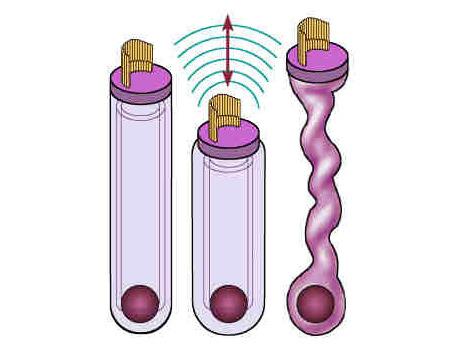
Figure 7 is a drawing of outer hair cells showing the effect of different internal pressures. The outer hair cell is divided in three parts. The top part is capped with a flat plate into which the stereocilia are inserted. The base of the cell is hemispheric. It contains the cell nucleus (round ball) and synaptic structures (not shown) for communicating with the central nervous system. The middle part of the cell is cylindrical in shape. The shape of the outer hair cell is maintained by a pressurized fluid core that pushes against an elastic wall. The wall is reinforced by additional layers of cytoskeletal material and membranes (shown in drawing by concentric cylinders). If the cell is at a normal pressure as on the left it will shorten and become fatter (middle) when there is an increase in intracellular fluid. When fluid is lost the cell elongates and the sides of the cell collapse (right) from the loss of pressure. Outer hair cells are isolated from the organ of Corti and kept alive to record their electromotility while recording their movements on video. During these experiments the outer hair cells will collapse, like the cell on the right, when exposed to large amounts of aspirin (equivalent to what you would have in your blood after taking 8 aspirin tablets). Aspirin has long been known to cause a reversible hearing loss and more recently it has been shown to block otoacoustic emissions. The experiments show that aspirin causes both effects by a direct action on the outer hair cell.
Most of the reinforcement is provided by two cytoskeletal proteins that are located immediately below the cell’s membrane. The stiffer of these is oriented circumferentially. These molecules serve the same function as the steel threads in steel belted radial tires. It is interesting that they are not directly attached to the cell membrane but instead appear directly bonded to another membranous structure that is located immediately under them. The membranes of this structure form what is equivalent to the inner tube of older tires. This "inner tube" lines the cylindrical portion of the cell and it is a structure that is found no where else in biology. It is called the subsurface cisterna and it is known to be essential for the full expression of electromotility. Manipulations that alter the subsurface cisterna reduce electromotility. The three layers that make up the side of the cylindrical portion of the outer hair cell are incredibly close together. The distance between the outside of the cell to the inside of the subsurface cisterna is much less than the wavelength of light. Up until recently the only way to see the layers was to use an electron microscope.
Next chapter: Active Hearing Improves Frequency Selectivity
Return to the table of contents of How the Ear Works


