
About the Lab
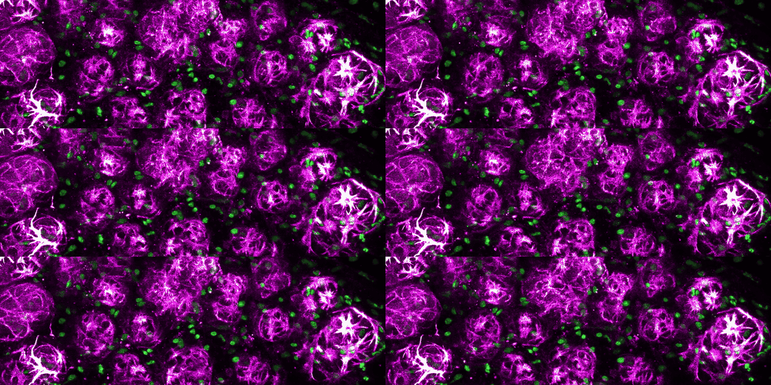
The Roarty Lab studies how cells within the breast communicate and coordinate their behavior, both during normal tissue building and during the breakdown of that order in cancer. Signaling between cells is only part of the story. What interests us is how those signals are translated into organized collective action, with cells taking on distinct roles, dividing labor, and moving as a unit. We treat tumors not as collections of independent cells but as communities, and the rules governing how those communities organize are central to explaining why breast cancers invade, spread, and resist treatment.
Our work is anchored in Wnt pathway biology, and spans mammary development, cell identity, and metastatic progression in triple-negative and other aggressive breast cancers.
Research Areas
Building and maintaining the mammary gland
The mammary gland is one of the few organs that develops largely after birth and remodels repeatedly across a lifetime. We investigate how canonical and alternative (β-catenin-independent) Wnt signals, including those routed through Ror-family receptors, are integrated within epithelial subsets to direct branching, position cells correctly within the tissue, and sustain the gland at steady state. A central question is how a single pathway achieves such specific outcomes depending on which cell receives the signal and when.
Cell identity, fate, and plasticity
Cells in the breast are not locked into fixed roles. We examine how epithelial cells acquire, maintain, and lose their identities, and how the plasticity that serves normal tissue becomes a liability once it is redeployed in cancer. This work addresses how heterogeneity within a tumor arises, how transitional and hybrid cell states are stabilized, and what those states contribute to progression.
Tumor ecosystems, adaptation, and metastatic vulnerability
Metastasis is rarely the work of one cell type acting alone. We treat the tumor as an ecosystem, in which subpopulations of cancer cells coordinate with one another and with stromal, immune, and vascular neighbors within a supporting matrix. What makes this ecosystem formidable is its adaptability. As cells leave the primary site and encounter the very different niche of a distant organ, the collective reorganizes itself: roles are reassigned, dependencies shift, and the group reconstitutes a functioning community in foreign tissue. Following that process from the breast outward, with an emphasis on the lung, we ask which coordinated behaviors and which niche interactions the ecosystem cannot do without. Those dependencies are the vulnerabilities we aim to target.
Approaches and Technologies
Our model systems include three-dimensional organoid culture, genetically engineered and syngeneic mouse models, patient-derived xenografts, and primary patient specimens. We visualize these systems by confocal and second harmonic generation microscopy, along with live imaging of tissue dynamics. To resolve the cell states and interactions underlying what we observe, we apply single-cell and spatial transcriptomics, flow cytometry, and additional multi-omics approaches.
The Lab
Our team is listed on the Lab Members page.