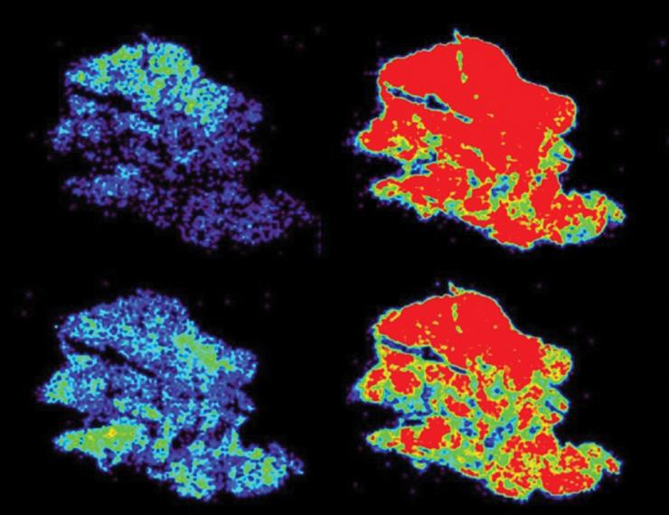
A major strength of our research team lies in advancing mass spectrometry imaging technologies for spatially resolved metabolic profiling of cancer tissues. We have developed and refined multiple approaches using desorption electrospray ionization (DESI) mass spectrometry (MS) imaging to enhance the specificity and depth of detection of hundreds of metabolites and lipids directly from human tissue sections. These methodological advances have enabled us to identify novel metabolic signatures and biomarkers that are diagnostic, prognostic, and predictive of treatment response across a range of diseases. One notable example is our work characterizing the metabolic composition of oncocytic thyroid and kidney tumors. Using DESI-MS imaging, we captured metabolomic information from distinct tumor regions within the heterogeneous tumor microenvironment. This analysis revealed that oncolytic tumors exhibit markedly altered cardiolipin profiles, including previously unreported oxidized cardiolipin species that may serve as therapeutic targets. Additionally, we have applied DESI-MS imaging to distinguish metabolic differences between cancerous and normal tissues, identifying molecular markers associated with tumor aggressiveness and chemotherapy response.
Our ongoing funded research continues to expand these applications, including:
- Imaging pediatric tumor tissues for disease stratification
- Identification of metabolic indicators of response to immunotherapies in human tissues (optimizing DESI-MS workflows for rapid detection of 9p21 loss, a pan-cancer genomic alteration associated with immunotherapy resistance)
- Molecular imaging of endometriosis and other complex diseases
Together, these efforts highlight the significant potential of MS imaging for biomarker discovery and for improving our understanding of disease biology and treatment response.