Neuroscientists create ultra-detailed map of brain region that controls movement
Hundreds of neuroscientists from across the globe, including researchers from the Tolias Lab at Baylor College of Medicine, have built a “parts list” of the motor cortex, mapping in exquisite detail the neurons and cells involved in a region of the mouse, monkey and human brain that controls movement. This first-of-its-kind atlas of a brain region lays the groundwork to map the whole brain and better understand brain diseases.
The large consortium of researchers is part of the BRAIN Initiative Cell Census Network (BICCN) brought together by the National Institutes of Health’s BRAIN Initiative.
The Atlas
The atlas described in a special package of 17 articles including a single flagship paper that describes the entire atlas, will be published today in the journal Nature. This includes a core-companion article describing research led by Dr. Andreas Tolias, professor, Brown Foundation Endowed Chair of Neuroscience and director of the Center for Neuroscience and Artificial Intelligence at Baylor.
The atlas is described as the first step to mapping the entire mammalian brain that will help researchers better understand brain diseases such as ALS, Parkinson’s disease and others that attack the neurons controlling movement.
The massive BRAIN Initiative-funded collaboration combined more than a dozen different techniques to define brain “cell types” across three different species of mammals and is one of the most comprehensive and detailed maps of any part of the mammalian brain. The researchers classified the millions of neurons and other kinds of brain cells present in the motor cortex into many different cell-type categories — the actual number of different brain cell types in this region depends on how they are being measured, but ranges from several dozen to more than 100.
“An integrated multi-modal atlas of cell types of the brain can be thought of as a cell-type Rosetta stone that relates genetic and phenotypic information. We believe this can provide a roadmap to understanding the logic behind the large diversity of cell types we see in the brain. It will help us decipher their function to ultimately understand how behaviors like perception and cognition arise from the wetware of the brain. These are some of the great frontiers of science.” said Tolias.
Tolias Lab core companion research
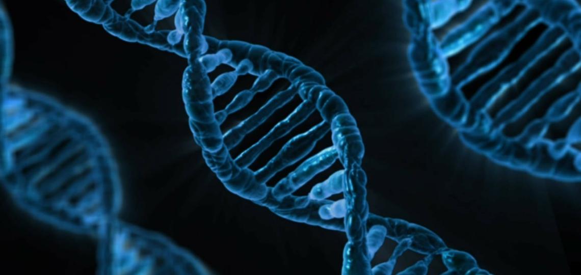
Tolias’ work, Phenotypic variation of transcriptomic cell types in mouse motor cortex, was a collaboration among his group, Dr. Philipp Berens from the University of Tübingen in Germany and Dr. Rickard Sandberg from the Karolinska Institute in Stockholm, Sweden. They describe the use of a Patch-seq, a technique they developed several years ago, to collect a large multimodal database including genetic, anatomical and physiological information from single cells in the mouse motor cortex.
They created a “tree of cell types” to get a better view of how neurons in the motor cortex are related to one another. This hierarchy of cell types shows one of the most detailed and complete characterizations of the diversity of neural types in the brain. It shows distinct, non-overlapping branches at the level of families. Within each family, neurons show continuous changes in their genetic, anatomical and physiological features, and all three features within a family are correlated. Read more about this research and collaborators on this project here.
BICCN
The NIH launched the BICCN in 2017, awarding nine collaborative network grants.
Like a population census, the cell census aims to catalog all different types of brain cells, their properties, their relative proportions and their physical addresses to get a picture of the cell populations that together form our brains. Knowing the “normal” brain’s cellular makeup is a key step to understanding what goes wrong in disease.
The researchers picked the primary motor cortex in part because it’s similar across all mammalian species — while humans, monkeys and mice have many differences between our brains, the way we control movement is very similar — and because it’s representative of the neocortex, the outermost shell of the mammalian brain that not only integrates sensory and motor information but also gives rise to our perception and other complex cognitive functions.
The studies required not only collaboration among researchers to design and execute the experiments, but also coordination and public sharing of the data that resulted from the atlas project and other projects under the BICCN. The Brain Cell Data Center, or BCDC, is headquartered at the Allen Institute. The data center, led by Allen Institute for Brain Science Investigator Dr. Michael Hawrylycz, helps to organize the BICCN consortium and provides a single point of access to the study’s data-archiving centers across the country.
Full funding information can be found in both Nature publications.