


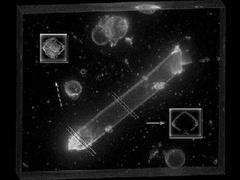
The cell is lying in bathing medium surrounded by assorted cellular debris. A 30 µm layer of the medium is rendered from 60 optical sections collected at 500 nm intervals. The images were deconvolved by MicroTome (VayTek, Inc.) and 3D rendered with VoxBlast (VayTek, Inc.). The plasma membrane has been stained with the fluorescent lipid analogue di-8-ANEPPS. The boxes show slices from the base and the middle of the cell. The slices were digitally extracted, further enhanced and rotated. Dye has entered the base of the cell because of synaptic related pinocytosis. In contrast, the dye is only present on the plasma membrane in the central portion of the cell. Fluorescent recovery after photobleaching was used to measure lipid mobility in the central region. Lipid mobility was found to be voltage- and tension-dependent. [Sections collected by Hong-Bo Zhao, image rendered by John Kesterson, composition of final image by Mas Takashima].
Learn more about: Hearing and Hair Cells | How the Ear Works