




Below you will find details on the following lab projects:
- Project 1: How to Restore a Functional Biliary System
- Project 2: N-Glycosylation and De-N-Glycosylation in Health and Disease
- Project 3: O-Glycosylation & Muscular Dystrophy
Project 1: Mechanistic and Preclinical Studies on Biliary Development, with a Focus on Alagille Syndrome
Background and Unmet Need
The biliary system plays essential roles in mammalian physiology, including excretion of waste products from the liver and secretion of bile acids to aid gut fat digestion. Not surprisingly, intrahepatic bile duct (BD) paucity—defined as a reduced number of BDs per portal vein—underlies a number of cholestatic disorders with major adverse consequences. The syndromic form of BD paucity is seen in Alagille syndrome (ALGS), a multisystem developmental disorder caused by autosomal dominant mutations in JAG1 (95%) or NOTCH2 (1-2%), which encode key components of the Notch signaling pathway. BD paucity and its consequences often lead to cholestatic phenotypes (like severe itching) and end-stage liver disease in patients with ALGS. The only strategy to restore a functional biliary system in these diseases is liver transplantation; in fact, ALGS is the leading genetic indication for pediatric liver transplantation in the US. However, liver transplantation poses a significant challenge to this patient population due to cardiovascular and renal abnormalities in some patients, liver donor shortage, and long-term adverse effects of immunosuppression, especially in children. Unfortunately, there is no FDA-approved therapy to enhance BD development in ALGS or any other disease with BD paucity.
Key Discoveries
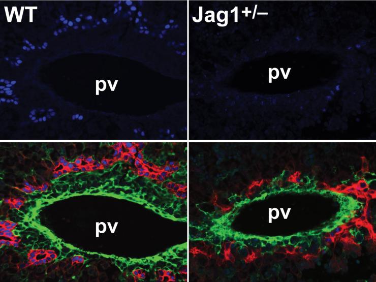
To address this unmet need, we first showed that on a pure C57BL/6 background, mice heterozygous for Jag1 (Jag1+/–) are a genetically representative mouse model for ALGS. Our subsequent identification of Sox9 as a dosage-sensitive enhancer of the Jag1+/– liver phenotypes allowed us to establish additional ALGS models with severe liver pathology. Using these models, we identified two dosage-sensitive genetic suppressors of the disease phenotypes: Poglut1, which encodes a glycosyltransferase, and Sox4, which encodes a transcription factor. Preclinical therapeutic studies in ALGS mice with moderate to severe bile duct paucity showed that antisense oligonucleotide (ASO)-mediated Poglut1 knockdown or adeno-associated virus (AAV)-mediated Sox4 knockdown can change the course of the disease by enhancing biliary tree development. Of particular interest were the observations that a single injection of our anti-Sox4 AAV in one-day-old (P1) mice led to long-term restoration of the biliary tree well into adulthood and that injection of this AAV at P15, when the mice already exhibit liver necrosis, was still able to enhance biliary tree development and rescue the liver phenotypes.
Current and Future Work
We are currently examining the molecular mechanisms underlying the rescue of the ALGS liver phenotypes by targeting each of these modifiers. Omics approaches are helping us elucidate the contributions of these genetic modifiers to biliary development and to identify cell type-specific responses to their knockdown that mediate the rescue of the ALGS liver phenotypes. Additional preclinical safety and efficacy studies aim to advance these strategies towards clinical trials. Another area of interest is to use these models to identify potential markers of disease severity and therapeutic response. Finally, while the focus of our work in this area has been ALGS, we are considering the possibility that the genetic modifiers identified in our ALGS studies might affect the phenotypes in other cholestatic diseases as well.
Collaborators and Funding
We gratefully acknowledge collaborations with the groups of Drs. Stacey Huppert and Nathan Salomonis (CCHMC), Jun Xie and Guangping Gao (UMass), Paymaan Jafar-Nejad and Frank Rigo (Ionis Pharmaceuticals), Saul Karpen (VCU), Bob Haltiwanger (UGA), Milton Finegold (BCM, TCH), and Neda Zarrin-Khameh (BCM, Ben Taub Hospital). Our work in this area has been generously supported by Baylor College of Medicine, the NIH, March of Dimes, The Medical Foundation (representing a donor advised fund), Texas Medical Center Digestive Disease Center, BCM Innovation Institute, and The Harrington Discovery Institute.
Project 2: The Roles of N-Glycosylation and De-N-Glycosylation in Animal Development and Human Developmental Disorders
Background and Unmet Need
Addition of carbohydrates to proteins (protein glycosylation) is among the most common post-translational modifications in nature, with important roles in development, growth and organismal physiology. About 3% of human proteins are involved in synthesis, degradation or recognition of glycans. Not surprisingly, over 160 human diseases—collectively called Congenital Disorders of Glycosylation or CDGs—are caused by pathogenic variants in the glycosylation machinery. These disorders primarily affect neonates and children, and present with a wide range of congenital anomalies affecting the nervous system, gastrointestinal tract, and the immune system among others, pointing to important links between glycosylation pathways and human developmental disorders. Importantly, an approved therapy only exists for ~10% of CDGs, highlighting the need for a better understanding of the mechanisms underlying the phenotypes caused by glycosylation gene mutations.
Key Discoveries
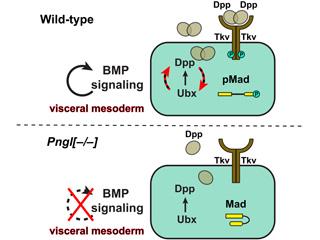
One of the most common forms of protein glycosylation is N-glycosylation, which plays broad roles in protein folding, trafficking, and ligand-receptor interactions, to name a few. So far, over 90 CDGs have been identified with mutations in N-glyco¬sylation machinery genes. Our work in this area has primarily focused on NGLY1, a cytosolic enzyme capable of removing N-glycans from misfolded N-glycoproteins during endoplasmic reticulum-associated degradation (ERAD). Recessive mutations in NGLY1 lead to a multisystem developmental disorder called Congenital Disorder of Deglycosylation 1 (CDDG1), which shows many phenotypic similarities to CDGs affecting N-glycosylation, along with some distinct features. Using Drosophila intestine as a model, together with transgenic mice as well as fibroblasts obtained from mutant mice and patients, we have identified several signaling pathways and processes regulated by NGLY1-mediated de-N-glycosylation. We reported that NGLY1 de-N-glycosylates the BMP4 ligand and its Drosophila homolog Dpp to promote BMP signaling in a tissue-specific manner. Importantly, our work suggests that NGLY1 is recruited to the cytosolic surface of the ER to promote the retrotranslocation of misfolded BMP4/Dpp from ER to the cytosol. We also found that NGLY1 regulates the expression of the major metabolic sensor AMPKα and promotes AMPK signaling in both flies and mammals. We have identified a critical role for NGLY1 in the regulation of gut barrier function, innate immunity, and lipid homeostasis in Drosophila.
Current and Future Work
Current work in this area aims to broaden the impact of these observations by investigating select components of the N-glycosylation machinery for their overlapping and divergent roles compared to NGLY1. These efforts have recently led to the identification of a novel evolutionarily conserved role for a component of the oligosaccharyl-transferase complex called TUSC3 as a dosage-sensitive gatekeeper between secretion versus degradation for BMP4 molecules. Another area of interest is to elucidate the roles of N-glycosylation and de-N-glycosylation in gut barrier integrity, innate immune response, and host-microbiota interactions. We employ complementary Drosophila and mammalian model systems to investigate these questions.
Collaborators and Funding
We gratefully acknowledge collaborations with the research groups of Drs. Tadashi Suzuki (RIKEN, Japan), Thomas Vaccari (U Milan), Tina Cowan (Stanford), Lars Steinmetz (Stanford & EMBL), Cat Lutz (JAX), Markus Affolter (Biozentrum, Switzerland), Antonio Galeone (CNR, Italy), and Hud Freeze (Sanford Burnham Prebys). Our work in this area has been generously supported by Baylor College of Medicine, the NIH, Mizutani Foundation for Glycoscience, and the Grace Science Foundation.
Project 3: O-Linked Glycosylation: From Drosophila Notch signaling to Human Muscular Dystrophy
Background
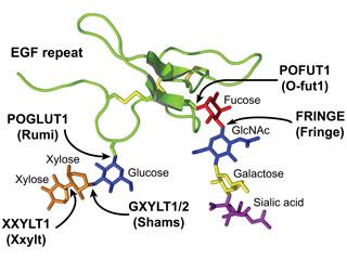
O-linked glycosylation of epidermal growth factor-like (EGF) repeats is a post-translational modification affecting over a hundred human proteins, with important roles in development and disease. One such modification is the addition of an O-linked glucose residue to EGF repeats with a specific consensus sequence. O-glucose can be extended by the addition of one or two xylose residues. The presence of xylose-xylose-glucose-O glycans on a number of proteins was discovered in the 1980s, but the enzymes responsible for this modification and their functional significance remained unknown for almost two decades.
Key Discoveries
In a forward genetic screen for Notch pathway modifiers in Drosophila, we identified POGLUT1 (encoded by rumi in flies), the enzyme responsible for adding O-linked glucose to EGF repeats in extracellular and secreted proteins in animals. We showed that POGLUT1 regulates fly Notch signaling in a temperature-sensitive manner and found that by altering the number of O-glucose residues on Notch, we can control the strength of Notch signaling. Genetic and cell culture experiments indicated that human and mouse POGLUT1 also regulate Notch signaling, although differences exist in the way the enzyme regulates fly versus mammalian Notch pathway.
In collaboration with Paradas and Haltiwanger groups, we identified recessive POGLUT1 mutations as the cause of a new form of limb-girdle muscular dystrophy, LGMDR21 (also called LGMD-2Z), revealing that skeletal muscle is uniquely sensitive to reduced POGLUT1 activity. Using conditional mouse models combined with biochemical and cell-based assays, we demonstrated that POGLUT1 plays critical roles in muscle stem cell development and maintenance by regulating Notch signaling and PAX7 expression. We have also characterized the functional roles of xylose residues that extend O-glucose on Notch EGF repeats. We found that xylose addition opposes Drosophila Notch signaling in specific contexts and plays a novel regulatory role by specifically affecting trans-activation of Notch by Delta ligands from neighboring cells, without affecting cis-inhibition by ligands in the same cell. This mechanism ensures optimal pathway activation in Delta-dependent developmental contexts.
Current and Future Work
Ongoing collaborative studies aim to characterize the roles of xylose-modifying enzymes in mammalian development and Notch signaling and to further investigate POGLUT1's role in muscle development and maintenance.
Collaborators and Funding
We gratefully acknowledge collaborations with the research groups of Drs. Bob Haltiwanger (UGA), Hideyuki Takeuchi (U Shizuoka, Japan), the late Hans Bakker (U Hannover, Germany), Radbod Darabi (U Houston), Carmen Paradas (U Sevilla, Spain), Tetsuya Okajima (U Nagoya, Japan), Yang Hong (U Pittsburgh), Amanda Simcox (Ohio State U), Lan Zhou (Houston Methodist Research Institute), and Michael Elowitz (Caltech). Our work in this area has been generously supported by Baylor College of Medicine, the NIH, Mizutani Foundation for Glycoscience, and March of Dimes.