The Cochlea

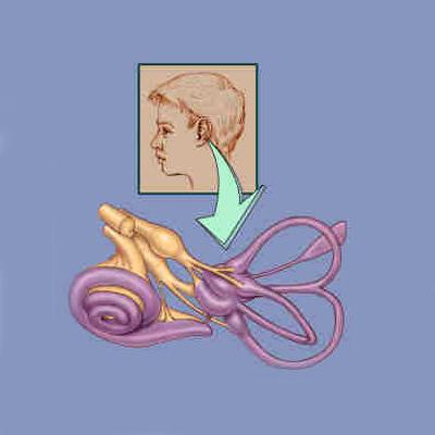
The hearing organ in mammals is a spiraling structure called the "cochlea" from the Greek word for snail. It spirals out from the saccule (one of the balance organs). There are two and a half turns in the human cochlea and if you were to unwind the cochlea it would stretch to nearly an inch in length. By winding itself in a spiral, the organ takes up less space.
The bony chambers (purple) of the inner ear form precise geometric shapes (see Figure 4). There are five sensory epithelia in the balance portion of the inner ear (one for each of the three semicircular canals and two in a chamber where the three canals come together). The nerve fibers (brown) can be seen on their way to the hair cells in the sensory epithelia. The nerve is made up of the neuronal projections that connect the hair cells with the brain and is called the eighth nerve because it is one of 12 nerves that come off the brain in the skull. The spiral shaped cochlea originates from one of the balance organs and contains the sensory epithelium for hearing.
The Organ of Corti - The Temple of Hearing

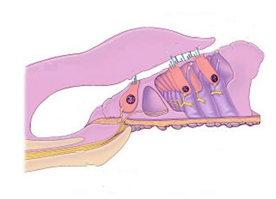
The sensory epithelium of the inner ear is called the organ of Corti after the Italian scientist who first described it. Its orderly rows of outer hair cells is unique among the organs of the body. Figure 5 shows a short section of the organ of Corti as it spirals in the cochlea. The organ of Corti is larger and the basilar membrane on which it sits is longer as it gets further away from the base of the cochlea. This difference in size is consistent with the fact that different frequencies of sound result in greater vibrations of the organ of Corti depending on where along the length of the cochlea you are measuring. The shorter, smaller structures near the base of the cochlea respond best to high frequencies, while the longer, larger structures near the top of cochlea respond best to low frequencies. This is similar to the organization of a piano where the longer, larger strings produce the lower frequency sounds.
The wide base of the cochlea from which this segment comes is towards the bottom of the page. The central axis of the spiraling cochlea is to the left of the drawing. Eighth nerve fibers pass through a bony shelf on their way to the hair cells (orange). The organ of Corti is made up of hair cells and supporting cells (purple and blue, respectively) that sit on a flexible basilar membrane which is anchored to the bony shelf on the left and a ligament (not shown) on the right. A single flask shaped inner hair cell is shown on the left and three rows of cylindrically shaped outer hair cells are seen on the right. The tips of the outer hair cell stereocilia are imbedded in a gelatinous mass called the tectorial membrane which lies on top of the organ of Corti and is secreted from cells (not shown) on the left. When sound is transmitted to the inner ear the organ of Corti begins to vibrate up and down. Since the basilar membrane is attached to bone and ligament at its two ends, the area of maximal vibration is near the third (furthest right) row of outer hair cells. The overlying tectorial membrane is not as flexible so the stereocilia are bent as the organ of Corti moves up and down against it. The electrical potential inside the hair cells changes as the stereocilia are bent.
Organization of Cells

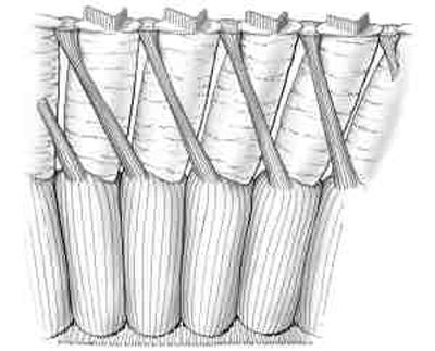
In no other organ in the body is it as easy to see the precise organization of the principal cells. The supporting cells of the organ of Corti are not found immediately adjacent to the outer hair cells so that for most of the length of these cylindrically shaped cells are surrounded by a relatively large fluid filled space (Figure 6 provides a view of a row of outer hair cells). The photogenic appearance of the organ of Corti has long been appreciated by the popular press. Pictures obtained with electron microscopes are routinely published showing the organ of Corti’s colonnade appearance. The three rows of columns are the outer hair cells and the organ is beautiful for the same reason that ancient Greek temples are beautiful. The space around the cells allows you to see their organization and appreciate their role in supporting the rest of the structure. In no other organ in the body do you find large fluid filled spaces around the principal cells. Neurons in the brain are surrounded by supporting cells. The cells in the muscles of the heart are close to one another. We now know that the spaces around the outer hair cell allow the cells to change their length during hearing.
Figure 6 is a view of a portion of the third row of outer hair cells. The view is what would be seen if you were looking towards the central axis of the cochlea and the most lateral set of supporting cells were removed. The organ of Corti is spiraling towards the top of the cochlea to the right. Stereocilia are on the top and radial fibers of the basilar membrane are seen on the bottom. The outer hair cells sit in a cup formed by a supporting cell. The supporting cells send out a narrow filament that angles towards the base of the cochlea. This unique structural organization means that the supporting cells touch the outer hair cells only at their top and bottom.
Next chapter: Inner and Outer Hair Cells
Return to the table of contents of How the Ear Works


