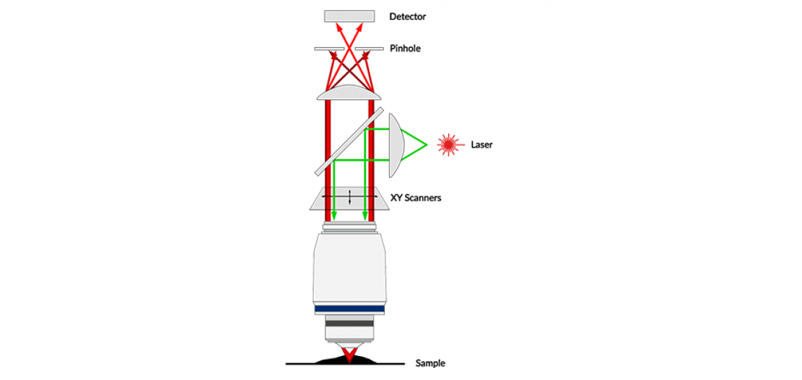
Confocal microscopy is an optical sectioning technique that utilizes fluorescence to generate a high resolution / high contrast image compared to traditional epifluorescence. These systems use a laser excitation source to illuminate the specimen in a single location (corresponds to a single pixel in a digital image). The emission from the fluorescence passes through a pinhole located in a conjugated focal plane that blocks signal from areas outside the plane of focus. The laser is then raster-scanned over the entire field of view, while fluorescence emission is recorded for a small axial volume at each location.

This technique leads to a significant improvement in axial resolution as well as contrast of the final image. Our confocal microscopes can be used to image a diverse range of biological specimens at sub-micron resolution. Many of our microscopes are equipped with incubation chambers that allow the regulation of temperature and CO² for live cell imaging over long periods of time. We also have a line scanning confocal microscope that allows one to image fluorescence at much higher speeds than the traditional point scanning approach.
See our ‘Available Instruments’ list (above right) for details about our confocal microscopes.
Optical Imaging & Vital Microscopy Core
Phone: (713) 798-6486
Email: oivm@bcm.edu


