Pneumonectomy is a surgical procedure to remove a lung.
It is performed for diffuse malignant pleural mesothelioma and other diffuse pleural cancers. Patients undergoing a pneumonectomy have normal liver and renal function and are typically under 70 years of age.
Tests such as ECG are performed to heart function and size, to check for valvular disease, and pulmonary hypertension.
Patients with pulmonary hypertension typically receive a temporary balloon occlusion of the main artery to the lung instead.
Before surgery is performed MRI and CT scans of the chest are done and in some cases exploratory surgery, to determine the extent of disease and to rule out mediastinal invasion and other contraindications.
Staging cervical mediastinoscopy and biopsy are also performed.
Surgery
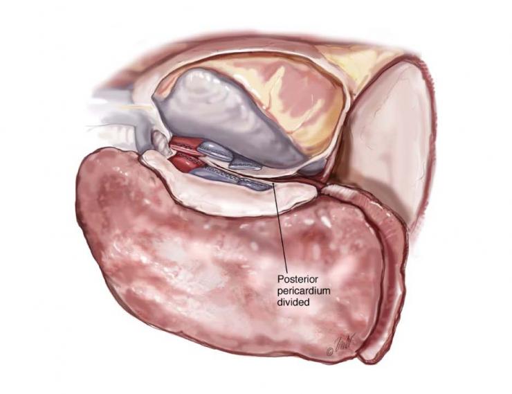
During the procedure the surgeon makes an incision and exposes of the space between the lung and the chest wall. Next they separate the tumor from the chest wall and then resect the lung, pleura, pericardium and diaphragm en bloc (in one piece) dividing the arteries, veins and bronchi that connect the lung to the heart. Lymph node dissection and reconstruction of the diaphragm and pericardium (membrane surrounding the heart) are then performed.
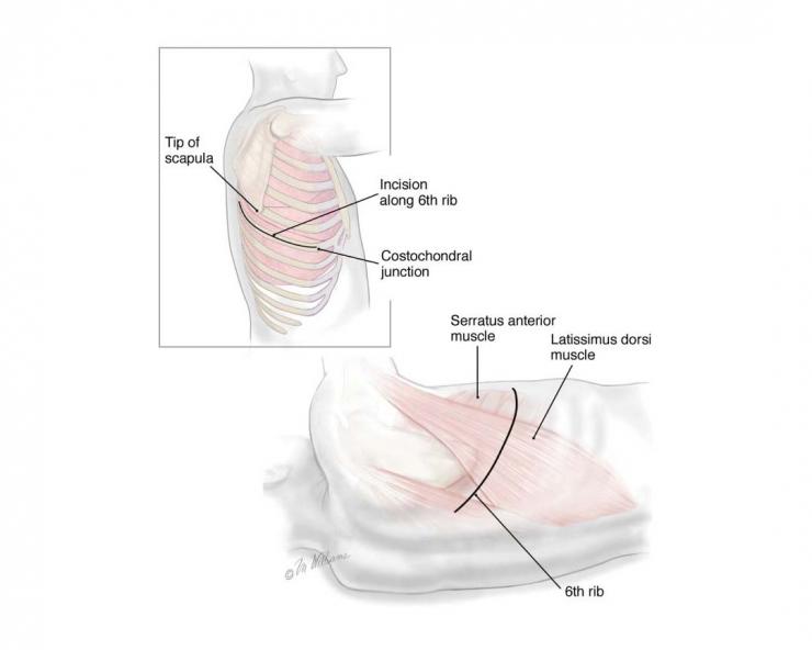
A thoracotomy is performed if the disease is contained in the chest cavity. The incision is started midway between the top of the shoulder blade and the spine and extends along the sixth rib. The large muscles along the side of the torso - serratus anterior and latissimus dorsi - are both divided. The sixth rib is removed.
Dissection
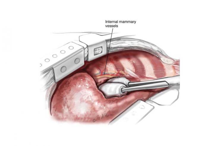
Two chest retractors are placed above and below the wound to spread and expose the area.
The surgeon will check for aortic and esophageal invasion. If found the procedure will be aborted.
The diaphragm (the muscle that raises and lowers during breathing) attachments to the chest wall are cauterized or pulled away. The peritoneum is bluntly dissected off the diaphragm. After the diaphragm is freed, the esophagus is dissected away from the resected tissues.
The right main bronchus is dissected close to the point it branches from one lung to another as with a bronchial stapler. With division of the bronchus, the en bloc resection is complete, and the tissue is removed from the chest.
Reconstruction of the Chest Wall
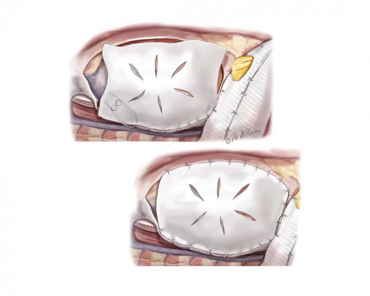
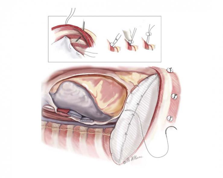
The diaphragm and pericardium are reconstructed with synthetic mesh material such as Gore-Tex. The mesh is formed to the chest wall with a fold in it to create a loose area at the center to reduce tension along the suture line and the chance of herniation. The mesh is sutured to the chest wall with nine sutures placed through the patch and between the ribs.
Reconstruction of Pericardium and Diaphragm
Each suture is passed through a plastic button and tied down onto button for added strength.
After the pericardial and diaphragmatic reconstruction is completed, an omental (fatty covering over the bowel) flap is sutured to the bronchial stump to cover and protect the area. Alternatively, an intercostal muscle or fat from around the heart may be used.
The thoracotomy is closed and rubber tube is placed into the pneumonectomy space and brought out of the incision.
After Surgery
Daily chest radiographs are obtained to assess mediastinal position and surveillance for infiltrates in the remaining lung. Any suggestion of pneumonia, clinically or biologically, is treated with antibiotics, chest physiotherapy, and bronchoscopy if needed.
Nasogastric tubes are used to decompress the stomach and to prevent aspiration. These are removed on day two after surgery. The red rubber catheter or chest tube is removed on day three after surgery.
References
Sugarbaker DJ, Bueno R, Krasna MJ, Mentzer SJ, Zellos L (eds). Adult Chest Surgery. New York: The McGraw-Hill Company, 2009.
Chang MY, Sugarbaker DJ. Extrapleural pneumonectomy for diffuse malignant pleural mesothelioma: Techniques and complications. Thorac Surg Clin. 14:523–30, 2004.
Garcia J, Richards W, Sugarbaker D. Surgical Treatment of Malignant Mesothelioma. Philadelphia, Lippincott-Raven, 1998.
Argote-Greene L, Chang M, Sugarbaker D. Extrapleural pneumonectomy for malignant pleural mesothelioma. In Multimedia Manual of Cardiothoracic Surgery (MMCTS). June 28, 2005.


 Credit