Our basic science and translational research focus falls under three broad categories.
Understanding the mechanisms that control intestinal development and homeostasis, and translating this knowledge into novel therapeutic approaches to treat diseases of the intestine such as IBD and colorectal cancer
Dr. Noah Shroyer’s laboratory elucidated roles for epithelial transcription factors such as Atoh1 (Math1), Gfi1, and Spdef in development and differentiation of the intestine. His laboratory has also translated these findings to human diseases, by showing that Atoh1 and its target Spdef are tumor suppressors that are frequently silenced in colon cancers, and that these genes are essential targets of Notch inhibitory drugs. In addition to these mechanistic studies, he and his team recently developed novel organ culture methods to direct differentiation of human pluripotent stem cells into intestinal tissue to study intestinal development and disease and used intestinal stem cell-derived organoids in quantitative assays to evaluate intestinal stem cell activity.
Understanding genomic and proteomic alterations associated with tumor progression of GI cancers
Dr. Ru Chen’s work in pancreatic cancer focuses on dissecting the signaling pathways and protein interaction networks that orchestrate tumor growth, invasion and metastasis. Some of these molecular alterations are currently under investigation for their utilities in clinical applications, such as: molecular imaging markers for identifying primary and metastatic pancreatic cancer, biomarkers for molecular diagnostics of pancreatic cancer and development of novel therapeutic targets. In addition, she is interested in a cytoskeletal protein called palladin. Her previous work found that overexpression of palladin gene (PALLD) in fibroblasts activated them into myofibroblasts, and enhanced cellular migration, invasion, and invadopodia-driven degradation of extracellular matrix. By understanding how PALLD promotes cancer development, her lab hopes to develop novel strategy directed at these mechanisms. Another focus of her lab is studying neoplastic progression of colitis-associated colorectal cancer. She found abnormal genomics and proteomics profiles in histologically normal appearing colon epithelial cells in chronic ulcerative colitis, and these molecular abnormalities could occur as a field defect in early tumorigenesis. Recently, her group is expanding to investigate gut microbiome at protein levels to identify microbial functions that are contributing to colorectal cancer and IBD.
Understanding the role of circadian hemostasis in cancer prevention and therapy
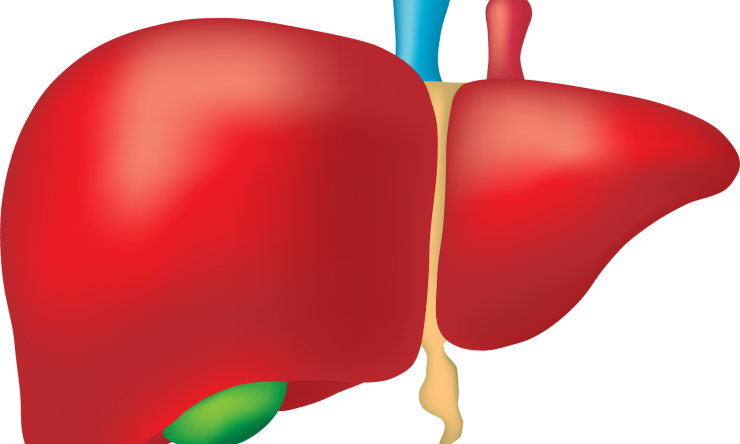
Evolutionary adaptation dictates that most physiological processes in mammals follow a circadian rhythm, which in all species studied is generated by an endogenous circadian clock. The circadian clock constantly couples internal physiology with external environmental cues to provide the best fitness. Recent studies have demonstrated that the prevalence of chronic circadian disruption in industrialized societies due to lifestyle changes significantly increases the risk of various modern-day diseases including metabolic syndromes and cancers. Dr. Loning Fu and her team discovered that disruption of the mammalian circadian clock increases the risk of various types of cancer. Their current studies are focused on the role of circadian homeostasis in the prevention and treatment of obesity and metabolic syndromes as well as obesity-related cancers, such as spontaneous hepatocellular carcinoma (HCC), which is previously considered a rare type of cancer in the Western countries but is expected to become a common cancer in the 21st century due to the prevalence of obesity and obesity-related liver metabolic disorders.
Understanding paligenosis: how mature cells revert to stem cells to fuel repair and (sometimes) cause pre-cancerous metaplasia and cancer
Professor Jason Mills laboratory studies how the tissues of the gastrointestinal system repair after damage. A large part of the repair process depends on “Cell Plasticity”, meaning the ability of cells to adapt to their environment by changing what they do. In fact, often, it is the mature, functional cells of an organ that are called upon to fix damage. To do that, they have to first stop their daily, physiological function (like secreting digestive enzymes) and retool their whole architecture to be like the young progenitor cells that help build the organ during the embryonic phases. It turns out that the mechanisms cells use to become embryonic-like progenitors are similar, no matter the cell or organ. Thus, the process whereby mature cells become progenitors again has been called Paligenosis. The Mills Lab studies the genes and cellular architecture changes that carry out paligenosis. For organs like the pancreas, paligenosis may be nearly the only way that the organ can repair itself, so regeneration after injury requires paligenosis. In organs of the luminal GI tract (esophagus, stomach, intestines), there are both normal, constitutively active, “professional” adult stem cells as well as cells capable of paligenosis. Harnessing paligenosis would help better invoke tissue repair. On the other hand, there is a downside to constantly recruiting, long-lived cells to become proliferative. Emerging evidence suggests that such cells don’t check for DNA damage and mutations as well as professional stem cells do. And, because, after injury they can return to the long-lived, mature state, they may warehouse whatever mutations they might have acquired during their moonlighting as stem cells, thereby increasing, with time, the overall mutation load of an organ and increasing the risk that eventually the paligenotic cells will not be able to return to their functional, mature state and will expand as an abnormal, precancerous, or frankly cancerous lesion. In short, the Mills Lab studies the origins of pre-cancerous lesions like metaplasia and cancer itself, which depend on the balance of stem and mature cell behavior.


