PLAIN RADIOGRAPHIC DIAGNOSIS OF CONGENITAL HEART DISEASE |
Contents | Previous Condition | Next Condition
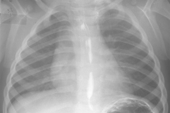
A. PA chest radiograph demonstrates a right sided aortic arch.
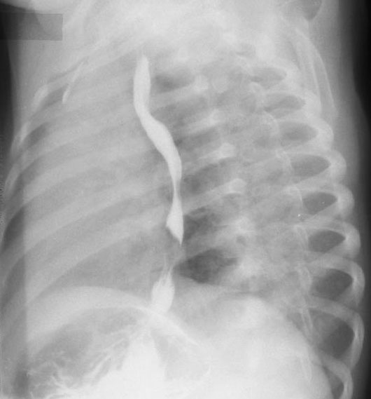
B. Left posterior oblique film demonstrates posterior indentation in the esophagram confirming the presence of a vascular ring. This infant had a right aortic arch with anomalous left subclavian arising from the descending aorta.
In some cases the left subclavian artery may arise from a retro-esophageal diverticulum (known as a Kommerell’s diverticulum) from the aorta. The ductus arises from the left side in the majority of cases, originating from the Kommerell’s diverticulum.
Hence the components of the vascular ring include the pretracheal ascending aorta, the right arch to the right of the esophagus and trachea, the left subclavian artery posterior to the esophagus and the ductus arteriosus or ligamentum on the left.