PLAIN RADIOGRAPHIC DIAGNOSIS OF CONGENITAL HEART DISEASE |
Contents | Previous Condition | Next Condition
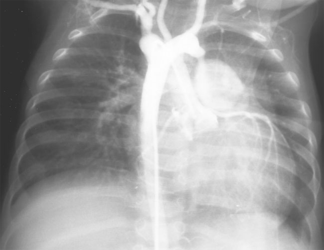
A,B. Cineangiograms demonstrate a double aortic arch with separate origins of carotid and subclavian arteries arising from each arch. The left arch appears to be dominant in this case with the descending aorta lying to the right of the spine.The main pulmonary artery can be seen filling from a ductus which arises from the left arch.
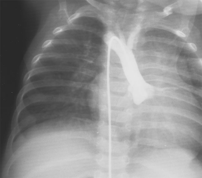
Double aortic arch is characterized by the persistence of both embryonic aortic arches. Each arch gives rise to an ipsilateral carotid and subclavian artery and there is no innominate artery. The ascending aorta divides into each arch anterior to the trachea and the larger arch crosses posterior to the esophagus and joins the contralateral arch to form a common descending aorta completing a vascular ring, which encircles both the trachea and esophagus. The ductus arteriosus usually arises from the left-sided arch. Both arches are usually patent and the right arch represents the dominant arch in some 70% of cases. The descending aorta usually descends on the side of the spine opposite to the dominant arch, usually the left side. Atresia of one arch is rare and if present normally involves the left arch.
Approximately 1 in 5 cases have an associated congenital heart defect including tetralogy of Fallot, transposition of the great arteries, ventricular septal defect or atrial septal defect. Rarely coarctation of one or other arch has been reported.