PLAIN RADIOGRAPHIC DIAGNOSIS OF CONGENITAL HEART DISEASE |
Contents | Previous Condition | Previous Image | Next Image | Next Condition
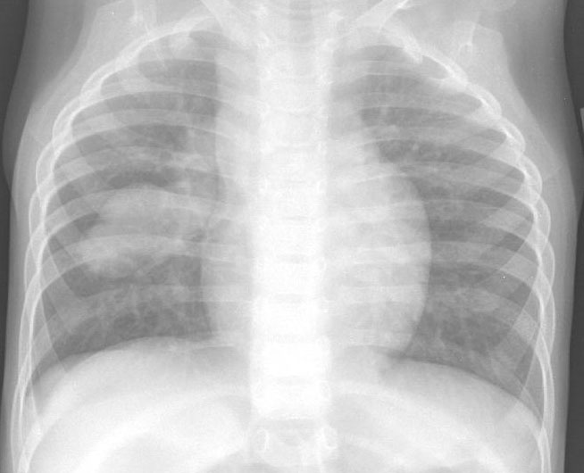
A. PA chest radiograph shows a globular shaped heart with widened superior mediastinum with an ovoid mass within the right middle lobe. This patient had anomalous left pulmonary veins which formed a venous confluence, crossed the midline and drained into a "pulmonary varix" prior to draining anomalously into the right superior vena cava. This patient also had heterotaxy syndrome with a common atrium.