PLAIN RADIOGRAPHIC DIAGNOSIS OF CONGENITAL HEART DISEASE |
Contents | Previous Condition | Next Condition
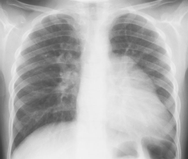
A. PA chest radiograph demonstrates prominent dilation of the left atrial appendage secondary to partial absence of the pericardium.
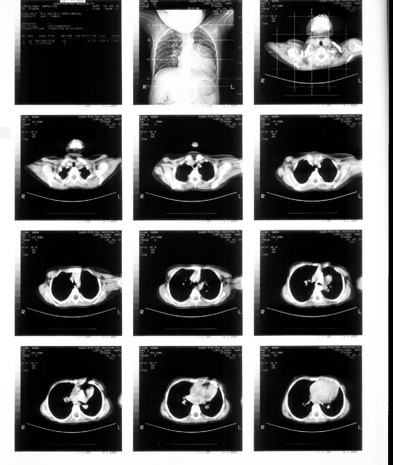
B. Noncontrast CT Thorax demonstrates dilation of the left atrium/left atrial appendage between frame 5 and 10.
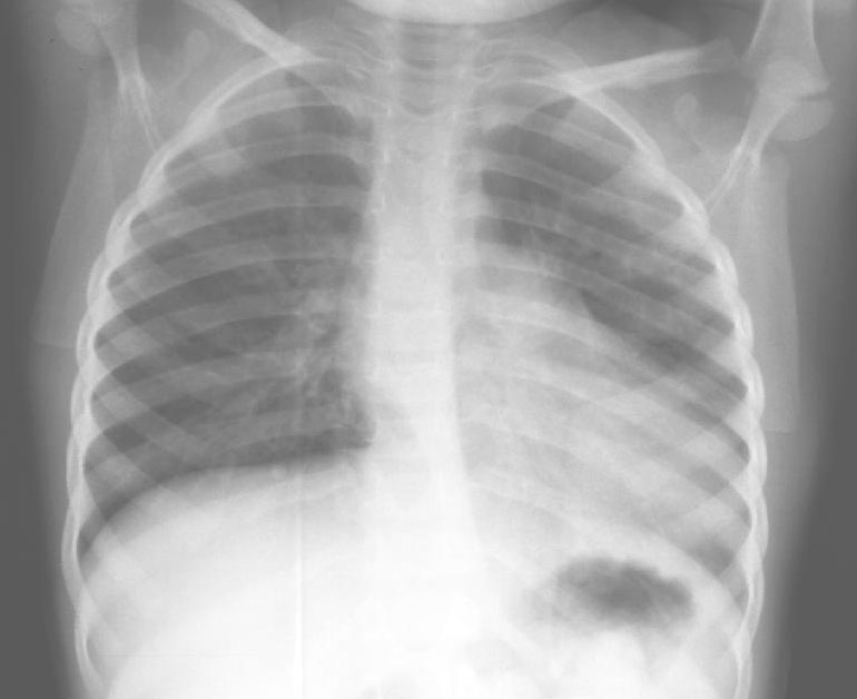
A. PA chest readiograph demonstrates cardiomegaly with prominent left atrial appendage secondary to complete absence of pericardium.
Columbus first described congenital absence of the pericardium in 1559. There is a male preponderance (3:1). Partial absence of the pericardium is more common than complete absence. The left pericardium is absent in 67% of cases with partial absence of the left pericardium being the commonest lesion. Right-sided lesions and bilateral absence are rare. In fetal life the common cardinal veins are responsible for blood supply to the pleuropericardial membrane wich eventually forms the pericardium. On the right side the common cardinal vein or right duct of Curvier persists as the right SVC, which ensures closure of the right pleuropericardial membrane. Early atrophy of the left duct of Curvier may give rise to left-sided pericardial defects including bronchogenic cysts, which are occasionally found in the pericardial space. In ľ cases of complete absence of the left pericardium a large pleural deficiency may allow herniation of the lung.