PLAIN RADIOGRAPHIC DIAGNOSIS OF CONGENITAL HEART DISEASE |
Contents | Previous Condition | Next Condition
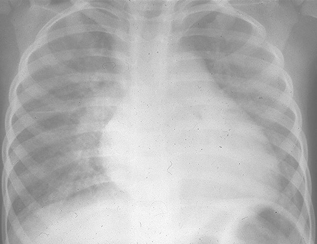
A. There is slight cardiomegaly. The pulmonary arterial markings are normal and there is an increase in the pulmonary veous pattern. Note also the faint Kerley B lines at the costophrenic angle.
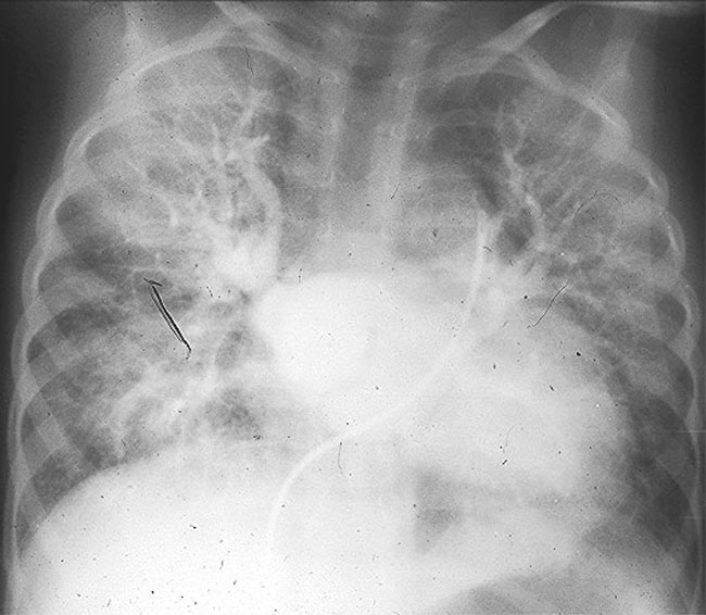
B. Angiocardiogram demonstrates the cor triatriatum septum within the left atrium.
Cor triatriatum (CT) is a rare congenital defect, which arises secondary to failure of incorporation of the common pulmonary vein into the leftatrium, resulting in an obstruction at the junction of the common pulmonary vein and the left atrium. This is most often a membranous obstruction with a single orifice through which pulmonary venous return can enter the left ventricle. Typically the left atrial appendage arises distal to the membrane in cor triatriatum compared to a proximal origin in supravalvular mitral ring.
Cor triatriatum occurs in approximately 4% children with congenital heart defects. It usually occurs in isolation although it may be associated with patent ductus arteriosus, left SVC to coronary sinus and VSD. There is an increased male prevalence.
CT presents with the same physiology as mitral stenosis. The natural history is the development of pulmonary venous hypertension and elevated pulmonary vascular resistance. If obstruction is severe in the absence of an atrial septal defect presentation occurs in infancy. The presence of an ASD may result in left to right shunting and minimize left atrial hypertension with presentation in later life.