PLAIN RADIOGRAPHIC DIAGNOSIS OF CONGENITAL HEART DISEASE |
Contents | Previous Condition | Next Condition
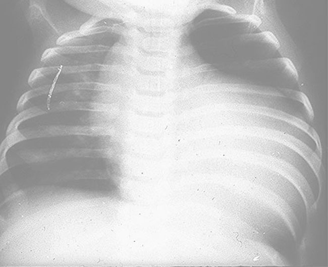
A. Chest radiograph.
Chest radiographs usually show cardiac dilation and pulmonary congestion similar to that seen in hypoplastic left ventricle. Interrupted aortic arch and truncus arteriosus are common findings in the Di George syndrome, a pure type of T-cell deficiency with absence or hypoplasia of the thymus and the parathyroid glands, as a result of failure of development of the third and fourth brachial arches. Neonatal tetany is usually the first clinical indication of this abnormality.