PLAIN RADIOGRAPHIC DIAGNOSIS OF CONGENITAL HEART DISEASE |
Contents | Previous Condition | Next Condition
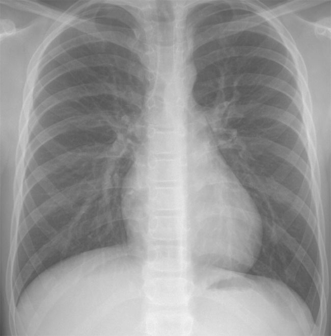
A. Posteroanterior (PA) chest radiograph demonstrates a slight increase in pulmonary arterial markings with a normal sized heart. The main pulmonary artery segment is convex.
ASDs have been recognised as early as the circulation was known with Leonardo da Vinci first documenting an ASD in a human postmortem.
An ASD is defined as any communication within the atrial septum which results in shunting between the atria.
These may be classified as:
Embryology: The atrial septum develops from a combination of the septum primum and septum secundum. The primum septum grows from the superior roof of the atrium towards the endocardial cushions (atrioventricular valves) from the fourth week of embryonic life. There is a gap or ostium primum between the inferior margin of the primum septum and the endocardial cushions. Before fusion of the primum septum with the endocardial cushions, a second opening, the ostium secundum, appears in the primum septum. Between week 5 and 6 of cardiogenesis, the septum secundum develops with 2 limbs, the inferior of which fuses with the lowest portion of the atrial septum joining with the endocardial cushions. There is an opening in the septum secundum called the foramen ovale, which lies immediately to the right of the septum primum and may result in a right to left communication between the atria. After birth, the two septa fuse in the region of the foramen ovale, however in 25-30% of normal hearts there is incomplete fusion of the flap of the septum primum and secundum with a patent foramen ovale with the potential for a right to left shunt and paradoxical embolism.
Genetics: Holt-Oram syndrome is associated with autosomal dominant inherited ASDs with absent or hypoplastic radii, and ECG findings of 1st degree AV block and RBBB. There is 100% penetrance with this disorder and approximately 40% of cases represent new mutations.