Klebsiella pneumoniae brain abscesses, with purulent meningoencephalitis, in a Patas monkey (Erythrocebus patas)
The patient was 13 months old, equivalent to about five human years, at the time he became ill in his quarters at the Houston Zoological Gardens. He developed focal left-sided seizures, accompanied by lateralizing signs on neurological examination suggesting the presence of a right hemispheric lesion. Urgent CT imaging identified multiple ring-enhancing lesions involving both cerebral hemispheres, with the largest lesion located in the right frontal lobe, corresponding to the patient's deficits. Several months before the patient's present illness, an antelope in the Houston Zoological Gardens had died from CNS toxoplasmosis. The patient was administered antibacterial antibiotics, seizure prophylaxis was initiated with phenobarbital, and a tapering dose of glucocorticoids was administered to minimize cerebral edema. As CNS involvement by toxoplasmosis may occur even in apparently immunocompetent primates, and given the history of a neighborhood exposure, the patient was empirically treated for CNS toxoplasmosis with sulfamethoxozole and pyrimethamine. Screens for immunodeficiency, including SIV (simian immunodeficiency virus), as well as screens for common primate viral illnesses (African green monkey herpesvirus and cytomegalovirus, in addition to human herpesvirus screens), were negative. A serologic assay for antibodies to Toxoplasma was also negative. Initially, the patient's symptoms improved, but his illness subsequently relapsed, with seizure recurrence. Despite additional antibiotic treatment, the patient died. Diagnosis was established by postmortem culture of purulent material from a brain abscess. No evidence of Klebsiella pneumonia, or other predisposition to cerebral abscess formation such as a valvular vegetation or pulmonary arteriovenous fistula, was found at necropsy.
Gram-negative bacterial meningitis, often arising as a complication of septicemia, is a major cause of infectious morbidity during the neonatal period and early infancy in human patients. Affected human children usually have an underlying condition predisposing to the development of gram-negative meningitis (e.g., Kaplan & Patrick, 1990). Although systemic and meningeal infection by Klebsiella pneumoniae is not unusual in human neonates and infants, brain abscess formation is quite uncommon in these age groups (Basu et al., 2001).
In contrast, Klebsiella pneumoniae infections are a major cause of morbidity and mortality in captive-bred, apparently immunocompetent nonhuman primates, even beyond the period of infancy. In some colonies, infection by Klebsiella pneumoniae is the leading cause of death (Postal et al., 1988). Transmission of organisms is believed to occur predominantly via fecal-oral routes, and colonization is believed to occur well before deep invasion of organisms in many cases (e.g., Kolodynski et al., 1989). Due to the substantial interest in maintaining healthy nonhuman primate colonies for zoological research, and for animal models of numerous human disorders, a capsular polysaccharide vaccine has been developed (e.g., Postal et al., 1988). Unfortunately, multidrug antibiotic resistance is common in organisms isolated from colonies of nonhuman primates, and the mortality associated with established Klebsiella pneumoniae infection remains quite high.
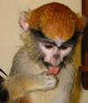
Photo courtesy of http://www.sierrasafarizoo.com
PRIMATES: Cercopithecidae; Genus Erythrocebus. The long limbs and relatively short feet and digits of the patas monkey, which lives on the African savannah, are adapted for fast running. The patas monkey, also known as the red monkey or hussar monkey due to its coloration, has been clocked at speeds in excess of 50 km/hr. Adult patas monkeys may weigh up to 13 kg, and may live as long as 20 years. Diet in the wild often consists of acacia gum, seeds, and insects (e.g., Isbell, 1998). These monkeys are often quite silent during usual activities, contrasting with many other monkey species, but in keeping with their tactics of concealment as a preferred defense against predators. Wild patas monkeys have been studied as models for foraging behavior, adaptations to terrestrial (as opposed to arboreal) locomotion, and primate social relationships. Studies of patas monkeys in captivity have advanced our knowledge of immunodeficiency viruses and herpesviruses in primate hosts, and our understanding of the relationships between cognition and cortical development.
We thank Maryanne E. Tocidlowski, D.V.M., of the Houston Zoological Gardens, for source material and for helpful comments on the presentation of the case. We also thank the staff of the Pediatric Neurology Service at the Texas Children's Hospital.
Dr. Kew's case presentation arose from an actual clinical consultation to the Pediatric Neurology Service at the Texas Children's Hospital. It therefore represents a real example of the range of clinical problems that could be encountered, albeit rarely, by an active clinician consulting on disorders affecting the nervous system. Several respondents commented on the brevity of the investigative workup, while others submitted diagnoses of various congenital syndromes that might account for the unusual description. We were surprised, however, that only one of the respondents raised the possibility that the patient might not be human.
To preserve the challenge of the case, we modified several details of the presentation as follows: (1) Scale bars were omitted from CT images and from biopsy (autopsy) material; (2) An explicit statement of the patient's species and living quarters was omitted; (3) Morphologic features (e.g., macrognathia) were generally described in relation to typical human proportions; and (4) The patient's tail was described as a prominent coccygeal region, and distinctive elements of coloration were omitted.
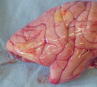
Gross brain, patient 64
We also scattered a number of clues throughout the description and the diagnostic studies. In brief, Dr. Kew described the patient as a 13-month old male with generalized hirsutism, bushy eyebrows, and a number of simian facial and limb features. A dramatic dissociation between gross motor and language development was evident in the history, typical for nonhuman primates, and less frequently encountered in human developmental disorders. The lateral skull film, available under the X-ray section, depicted the facial profile. The upper two CT images, presented under "CT: Head," hinted at an extremely unusual ratio of ocular diameter to brain size (for a human patient), as well as marked underdevelopment of the paranasal sinuses. We reasoned that in a patient presenting with suspected multifocal cerebral abscesses and a possible neighborhood exposure, many readers would check a serologic assay for Toxoplasma gondii (under "Serum/CSF Serologies") as part of the evaluation, and provided SIV (simian immunodeficiency virus) test results in that section. We regarded a report of this test (with the HIV screen "not done") as distinctly unusual for a human case, and an additional clue as to the nature of the patient. Finally, we reasoned that failure to diagnose the patient's symptomatic cerebral ring-enhancing lesions using noninvasive methods would prompt many readers to request a brain or lesional biopsy. Along with the biopsy (necropsy) results, we presented a view of the cortical surface of the left hemisphere, to convey the classic morphologic features of the simian brain.
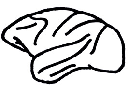
Adapted from Brodal A, Neurological Anatomy
We also scattered a number of clues throughout the description and the diagnostic studies. In brief, Dr. Kew described the patient as a 13-month old male with generalized hirsutism, bushy eyebrows, and a number of simian facial and limb features. A dramatic dissociation between gross motor and language development was evident in the history, typical for nonhuman primates, and less frequently encountered in human developmental disorders. The lateral skull film, available under the X-ray section, depicted the facial profile. The upper two CT images, presented under "CT: Head," hinted at an extremely unusual ratio of ocular diameter to brain size (for a human patient), as well as marked underdevelopment of the paranasal sinuses. We reasoned that in a patient presenting with suspected multifocal cerebral abscesses and a possible neighborhood exposure, many readers would check a serologic assay for Toxoplasma gondii (under "Serum/CSF Serologies") as part of the evaluation, and provided SIV (simian immunodeficiency virus) test results in that section. We regarded a report of this test (with the HIV screen "not done") as distinctly unusual for a human case, and an additional clue as to the nature of the patient. Finally, we reasoned that failure to diagnose the patient's symptomatic cerebral ring-enhancing lesions using noninvasive methods would prompt many readers to request a brain or lesional biopsy. Along with the biopsy (necropsy) results, we presented a view of the cortical surface of the left hemisphere, to convey the classic morphologic features of the simian brain.
Additional clues, embedded in the description and lab reports, are left to the interested reader. It is often said that a picture is worth a thousand words, emphasizing the difficulty of forming a detailed image from verbal description. We would note that this familiar aphorism is sometimes reversed: a few key words, a questioned assumption, or a unifying hypothesis may advance the understanding of a number of disparate pictures.
-- Dennis R. Mosier, M.D., Ph.D.
Email comments: