Spontaneous intracranial hypotension;
Secondary Arnold-Chiari malformation
Patient #28 is a young, healthy (though mildly obese) female who presented with new onset headaches and a normal neurological exam. Normally, this would suggest the possibility of migraine, tension headache, pseudotumor cerebri, or a mass lesion. The characteristics of the headache, however, are inconsistent with any of the major categories of headache syndromes. The pain was not well-localized, did not have a throbbing character, and was not associated with vomiting, visual changes, photophobia, or phonophobia. Furthermore, the headaches were clearly related to position, worsening upon standing and resolving upon assuming a supine position. These symptoms are opposite of those caused by increased intracranial pressure, and suggest orthostatic headaches due to intracranial hypotension. Certainly many headaches from other etiologies can be worsened with standing or movement (e.g. migraine or meningitis) but, in general, these do not completely resolve when recumbent and recur with standing. The differential diagnosis for isolated orthostatic headaches includes spontaneous intracranial hypotension, colloid cyst of the third ventricle and post-lumbar puncture headache.

Figure 1. A coronal T1-weighted image, post-gadolinium, before treatment: There is diffuse leptomeningeal enhancement throughout, including both hemispheres, basal dura, and tentorium cerebelli. The enhancement is smooth and symmetric, without nodular features. There is cerebellar tonsillar descent. There is no mass effect, mass lesion, hemorrhage, infarct, or parenchymal enhancement.
The most worrisome and confusing aspects of this case, however, were the findings of CSF pleocytosis and diffuse meningeal enhancement on MRI (see Figure 1). In fact, the patient was referred to our service for a possible meningeal biopsy because of the unexplained nature of her problem and these MRI findings. The numerous possibilities of diffuse meningeal enhancement include inflammatory, infectious, and neoplastic processes.
Meningeal enhancement is seen with meningeal carcinomatosis and lymphoma but the appearance is usually one of nodular enhancement, which may be focal or diffuse. Our patient had no evidence of blood cell dyscrasia on peripheral smear. Additionally this patient had no other symptoms or laboratory findings to suggest the presence of a malignancy.
Neurosarcoidosis can give a similar appearance but it is usually associated with cranial neuropathy (53%), parenchymal disease (48%), aseptic meningitis (22%), peripheral neuropathy (17%), myopathy (15%) and/or hydrocephalus (7%). There is usually some evidence of systemic disease (97%) and elevation of serum angiotensin enzyme (ACE) levels. None of these were present in our patient.
Lyme disease was considered. There is a triad of meningitis, radiculitis and neuritis without fever that is highly suggestive of this disease. Meningeal enhancement may or may not be seen, but is more often at nerve roots. Initially, patients have mild meningeal signs including headaches, myalgias, stiff neck and cranial nerve involvement. After several weeks patients may have cardiac conduction abnormalities, meningial signs, multiple cranial neuropathies, peripheral mononeuropathies and encephalopathy, as well as transitory erythematous blotchy rashes. The third stage typically involves development of a chronic arthritis. Our patient had none of these findings and her Lyme titers were negative.
Multiple infections, too numerous to be listed here, including viral, fungal and bacterial, are associated with meningeal gadolinium enhancement, usually with more advanced disease. In our patient, at least three separate sets of CSF cultures for bacteria, acid-fast bacteria, and fungus, were obtained, all of which were negative. Special studies for Histoplasmosis, Cryptococcus and Toxoplasmosis were, by report from the referring physician, also negative. Although meningitis may have many different presentations, a primary, essentially solitary, symptom of orthostatic headaches would be most unusual.
Other etiologies associated with meningeal enhancement include post-trauma, post craniotomy or craniectomy, subarachnoid hemorrhage and following intra-thecal chemotherapy which are not part of this patient's presentation.
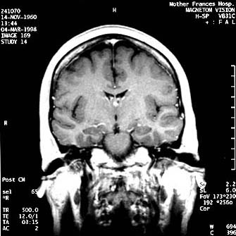
Figure 2. A coronal T1-weighted image, post-gadolinium, following treatment
The patient had no other systemic signs to suggest a more severe illness. Subsequent lumbar puncture confirmed the findings of a mild pleocytosis and elevated CSF protein. The first attempt at a lumbar puncture was unsuccessful, most likely due to the patient's low CSF pressure. A subsequent LP, performed under fluroscopic guidance, showed an opening pressure of 68 mm H2O, consistent with CNS hypotension and possible spontaneous CSF leak. The location of the CSF leak in this case is unclear. The initial MRI of the spine showed a suggestion of an anteriorly located extradural collection of CSF in low cervical-high thoracic region. Unfortunately the radionucliotide cysternogram was not confirmatory.
Based on the presumption of a spinal CSF leak, our patient was treated with a blood patch, using 15-20 cc of autologous blood injected into the posterior lumbar epidural space, after which the patient was placed in reverse Trendelenberg position lying on her stomach for two hours. Immediately following the procedure she was able to stand and walked without complaints. She has remained headache-free since that time. Repeat MRI shows complete resolution of the meningeal enhancement and loss of the cerebellar descent with restoration of the cerebellar tonsils to their normal anatomic position (see Figure 2).
Diffuse meningeal enhancement associated with chronic intracranial hypotension was first described in 1991.[1] Numerous authors have since described the same phenomenon.[2,3,4] The clinical characteristics are usually orthostatic headaches with severe pain upon standing or sitting that is slowly relieved by assuming a recumbent position. Associated findings include nausea, vomiting, photophobia, diploplia or blurred vision. Physical examination is generally unremarkable although unilateral or bilateral sixth nerve palsies and neck stiffness may occur. A singular characteristic of this condition is the large discrepancy between the clinical appearance of the patient and the MRI findings.
As previously described, patient's MRIs show diffuse, smooth thickening of the meningies, which enhance with gadolinium. Often there is a generalized sagging of the brain with downward displacement of the cerebellar tonsils giving the appearance of a Type I Arnold-Chiari malformation. In the series reported by Mokori et al, 100% of the patients had diffuse meningeal enhancement, 69% had evidence of subdural CSF collections and 62% showed a descent of the brain resembling the Arnold-Chiari malformation.[2]
The cause of the meningial thickening is not known although compensatory venous engorgement secondary to the chronically low CSF volume (the Monroe-Kellie rule, which states that CSF volume and CNS intracranial blood volume are inversely related) has been offered as one possible explanation.[3] The venous engorgement then causes both an appearance of thickened dura and a relative accumulation of the contrast media, gadolinium. Dural microvasculature does not contain the tight junctions of the arachnoid matter responsible, in part, for the blood brain barrier. This would allow for interstitial accumulation of contrast media.[3] In some patients with long-standing CSF hypotension there is also fibrosis of the meningies. Whatever the specific cause, most authors agree that the precipitating event is usually a spontaneous or iatrogenic CSF leak, e.g., post lumbar puncture, overdraining shunts, etc. CSF opening pressures are low (<40 mm H2O) in about 46% of patients. There is variable pleocytosis, usually modest, with occasionally elevated CSF protein. Otherwise CSF studies are normal.
Treatment consists of repairing the CSF leak, most often with 15-20 cc of an autologus blood patch. Resolution of orthostatic headache symptoms occurs within a few hours and the brain returns to normal anatomic position within a few days with loss of gadolinium enhancement.[2,3]
Email comments: