PLAIN RADIOGRAPHIC DIAGNOSIS OF CONGENITAL HEART DISEASE |
Contents | Previous Condition | Next Condition
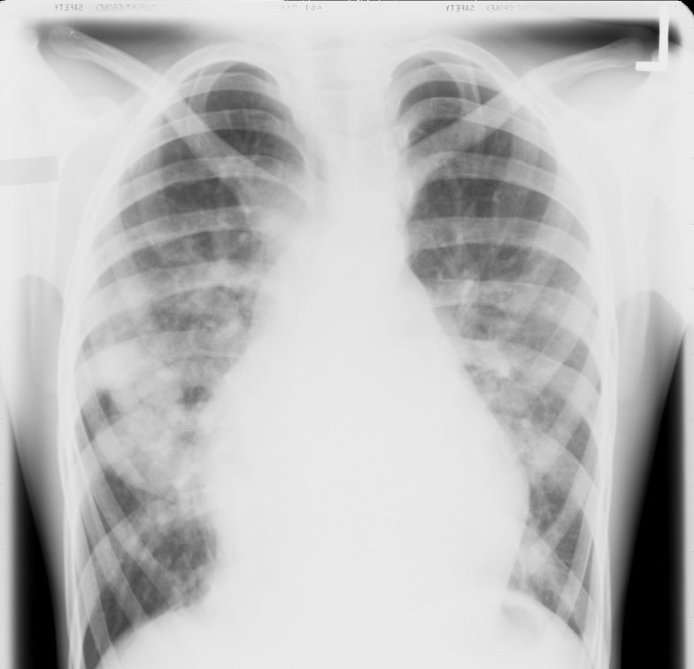
A. There is moderate cardiomegaly with prominence of the main pulmonary artery segment and the hilar vessels. There are large irregular sharply marginated densities in the right middle lobe which coalesce which represent pulmonary varices.
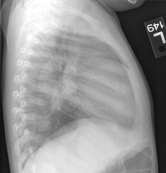
B. Lateral chest film demonstrates sharply demarcated ovoid variceal structures.
This patient had anomalous pulmonary venous drainage associated with variceal dilation and an atrial secundum defect. Pulmonary varices are also known as pulmonary "pyles".
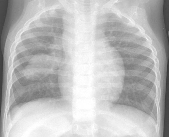
A. PA chest radiograph shows a globular shaped heart with widened superior mediastinum with an ovoid mass within the right middle lobe. This patient had anomalous left pulmonary veins which formed a venous confluence, crossed the midline and drained into a "pulmonary varix" prior to draining anomalously into the right superior vena cava. This patient also had heterotaxy syndrome with a common atrium.
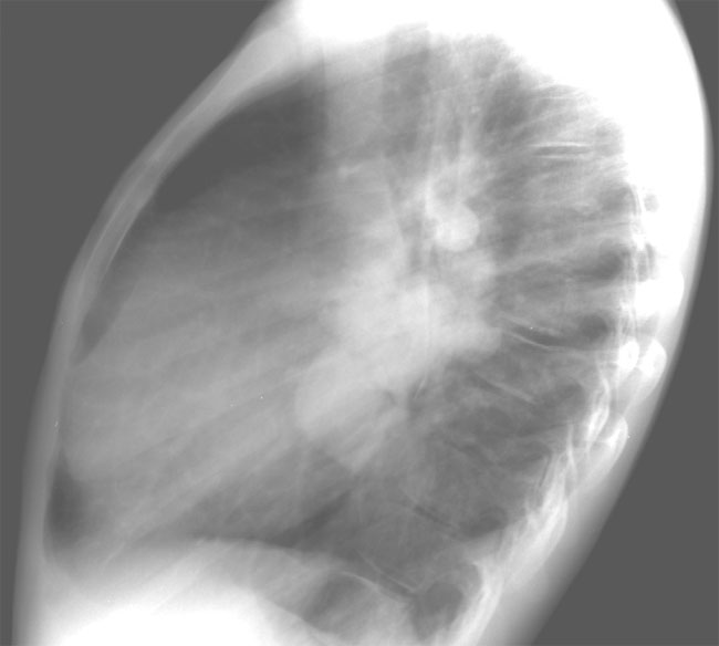
B. Lateral radiograph demonstrates the varix end-on with an ascending vertical vein draining into the right superior vena cava.