PLAIN RADIOGRAPHIC DIAGNOSIS OF CONGENITAL HEART DISEASE |
Contents | Previous Condition | Next Condition
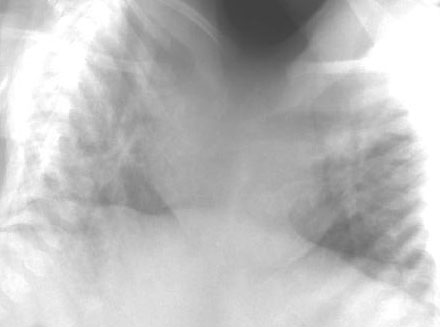
A. Plain film demonstrates thoracopagus conjoined twins with cardiac fusion but separate abdominal viscera.
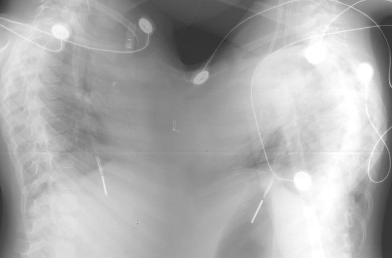
A. PA chest radiograph demonstrates thoraco-abdominus conjoined twins. These infants shared fused liver, intestine and ventricles. Twin A had tetralogy of Fallot; twin B had complete atrioventricular septal defect with infradiaphragmatic obstructed total anomalous pulmonary veins. There was a common atrium and bilateral superior vena cavae. There was ventricular fusion and separation was deemed unfeasible.