PLAIN RADIOGRAPHIC DIAGNOSIS OF CONGENITAL HEART DISEASE |
Contents | Previous Condition | Next Condition
A.

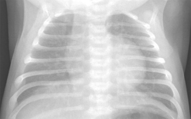
Tricuspid atresia represents the third commonest form of cyanotic congenital heart disease, and is characterized by complete absence of the communication between the right atrium and ventricle. This lesion occurs in approximately 1:15,000 live births.
Classification: There are three types classified according to the relationship of the great arteries: type I, normally related great arteries; type II, transposed great vessels, and type III, l-transposed great arteries. These types are subclassified according to the presence/size of associated ventricular septal defect and the presence or absence of pulmonary atresia. There is a obligatory atrial communication to enable right to left shunting and decompression of the right atrium.
Associations: There is a higher incidence of associated anomalies in type II (63%) than type I (18%). These include persistent left superior vena cava to coronary sinus (12-15%). Juxtaposition of the atrial appendages occur in approximately 10% of all cases and up to 40% of type II. Other associated defects include coarctation of the aorta (8%), patent ductus arteriosus (3%), and right aortic arch (3-8%). The left bundle branch originates early from the bundle of His, which is responsible for the high incidence of superior axis on ECG. The presence of juxtaposed atrial appendages is relevant to surgical palliation particularly if a modified atrio-pulmonary (Fontan) communication is to be performed.