PLAIN RADIOGRAPHIC DIAGNOSIS OF CONGENITAL HEART DISEASE |
Contents | Previous Condition | Next Condition
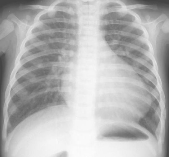
A. The heart is enlarged with a narrow "pedicle" giving the so called "egg on a string" appearance. The superior mediastinum appears narrow due to the antero-posterior relationship of the transposed great vessels and "radiologic-absence of the thymus".
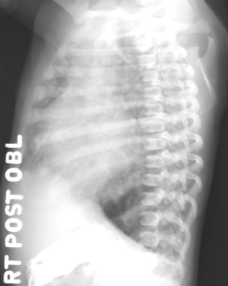
B. The stress of hypoxia in the newborn period is believed responsible for thymic regression. Right posterior oblique view demonstrates widening of structures in the superior mediastinum due to the anteroposterior relationship of the aorta and pulmonary artery.
D-TGA accounts for some 5% of all congenital heart defects. D-TGA describes patients in whom there is atrioventricular concordance with ventriculoarterial discordance i.e. the pulmonary artery arises from the left ventricle and the aorta arises from the right ventricle. Baillie is accredited with the first documented case in 1797. Prior to the introduction of atrial septectomy by Blalock and Hanlon in 1950, the mortality rate from transposition was as high as 90%. The management of these patients was further revolutionized in 1966 with the introduction of balloon atrial septostomy by Rashkind and Miller.
Pathology: Situs solitus is usually and present. In the right ventricle there is a muscular infundibulum separating the aortic and tricuspid valves, while in the left ventricle there is fibrous continuity between the leaflets of the pulmonary and mitral valves. The aorta is positioned anterior and generally to the right of pulmonary artery.
Embryology: Abnormal looping of the great vessels is believed to occur. In the normal heart a bilateral conus develops beneath both semilunar valves with progressive development of the subpulmonary conus and regression of the subaortic conus, resulting in the great vessels arising from the appropriate ventricle. In d-TGA the reverse occurs with progressive growth of the subaortic conus and regression of the subaortic conus resulting in transposition of the great arteries.
Associated lesions: These include VSDs (typically outlet or muscular septum defects) often with malalignment of the outlet septum which is deviated rightward, resulting in overriding of the pulmonary valve. If there is significant override this may represent Taussig-Bing anomaly (double outlet right ventricle with subpulmonary VSD). Other associated lesions include pulmonary stenosis (30%), patent ductus arteriosus, coarctation, coronary artery anomalies (the right and circumflex coronary arteries arising from the right sinus, the circumflex coronary from the right sinus and the right and left anterior descending coronaries from the left sinus, intramural variant and single coronary arteries).
Physiology: This lesion behaves as two separate circulations with mixing at the atrial, ventricular, or ductal level essential for survival. Patients with restrictive atrial shunting should undergo atrial septostomy to maximize atrial level mixing, and minimize acidosis prior to arterial switch operation. This may performed either by the bedside under echocardiographic guidance, or particularly in cases where the anatomy cannot be fully elucidated from echocardiography, in the cardiac catheterization laboratory