PLAIN RADIOGRAPHIC DIAGNOSIS OF CONGENITAL HEART DISEASE |
Contents | Previous Condition | Next Condition
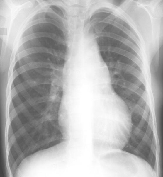
A. The heart is slightly enlarged, the main pulmonary artery convex, and the aortic arch prominent above the MPA. There are increased pulmonary vascular markings.
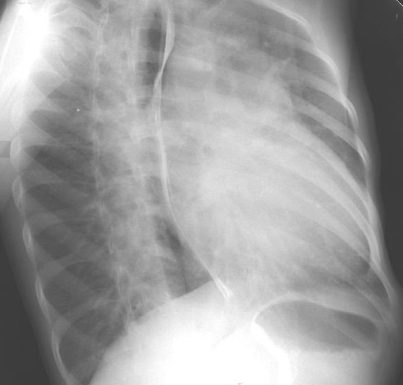
B. Right anterior oblique view demonstrates the esophagus to be indented by a large left aortic arch in addition to posterior displacement by the dilated left atrium.
Persistent patent ductus arteriosus occurs in 1 in 2500 to 1 in 5000 live births and represents between 9-12% of all congenital heart defects. There is a 2:1 female to male ratio.
Embryology: the ductus arteriosus originates from the distal portion of one of the sixth paired aortic arches and enables communication between the pulmonary artery and the descending aorta. In an infant with a normal left aortic arch, the ductus originates from the distal left sixth aortic arch and connects the main pulmonary artery to the left descending aorta distal to the origin of the left subclavian artery. With a right aortic arch the ductus normally still arises from the distal left sixth arch along with a remnant of the left dorsal aorta (Kommerell’s diverticulum), either as an isolated structure or in association with an anomalous left subclavian artery; in both these cases the ductus or its ligamentum passes behind the trachea and esophagus and creates a vascular ring. Rarely in infants with a right aortic arch, the ductus arises from the distal right sixth aortic arch and connects the right pulmonary artery to the right descending aorta distal to the origin of the right subclavian.
Associations: Infants of prematurity, patients exposed to hypoxemia, birth at high altitudes, female sex and exposure to rubella particularly in the first 4 weeks of pregnancy are all associated with an increased incidence.
Physical examination: The classical findings are diagnostic. The pulses are bounding. The blood pressure has a wide pulse presssure. The apex beat may be displaced. There is a LV impulse. The second heart sound is loud and widely split with a "slap-sail" quality. The murmur is characteristically continuous or machinery like in quality. The diastolic component may disappear as one move down the sternum, particularly in smaller infants.
Complications: These include congestive cardiac failure, bacterial endocarditis, aneurysmal dilation of the ductus and potentially the development of pulmonary hypertension.
Differential diagnosis: This includes the innocent venous hum (obliterated with pressure over the inferior neck), aortopulmonary window, truncus arteriosus, absent pulmonary valve syndrome, VSD with aortic regurgitation, ruptured sinus of Valsalva with fistula, systemic and pulmonary arteriovenous fistula, aortopulmonary collaterals, coronary artery fistula and peripheral branch pulmonary stenosis.