PLAIN RADIOGRAPHIC DIAGNOSIS OF CONGENITAL HEART DISEASE |
Contents | Previous Condition | Previous Image | Next Image | Next Condition
Because of the variations of the mediastinal silhouette, as a result of thymic size and variations in the respirations, the descriptions of the radiographic anatomy does not become consistent until later childhood. Admittedly, echocardiography has replaced oblique views of the chest in the evaluation of cardiac chamber enlargement. However, the radiographic anatomy should be understood.
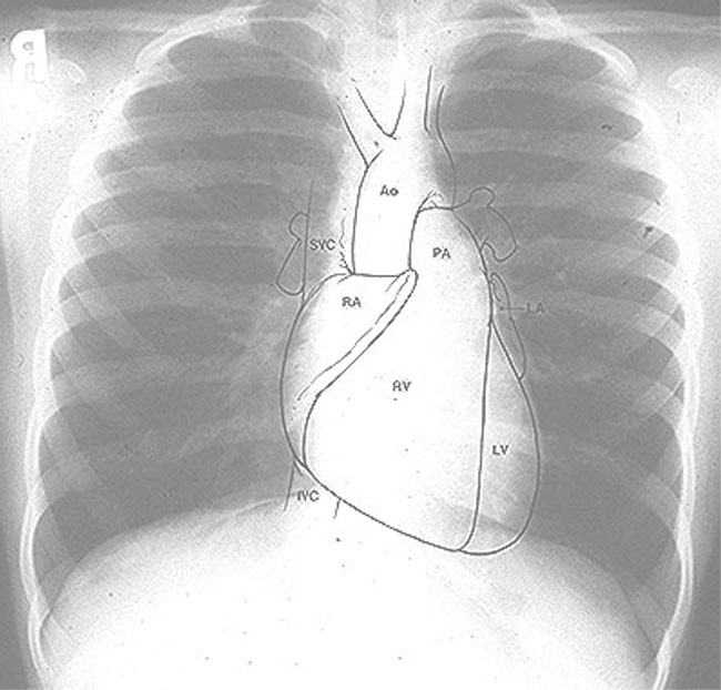
In the frontal projection, the right cardiac border is the right atrium and the left cardiac border is normally the outflow tract of the left ventricle. Above the left ventricular convexity, there is a normal prominence of the undivided portion of the pulmonary artery. The convexity above this is the aortic arch which displaces the trachea slightly to the right. In spite of anatomic textbooks, the left atrial appendage rarely contributes to the left heart silhouette.
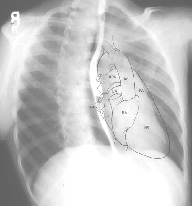
In the right anterior oblique view, the anterior chamber is the right ventricle, and the convexity above it is the pulmonary artery; posteriorly, the inferior shadow is the right atrium, and above it lies the left atrium. Enlargement of the left atrium will displace the esophagus (outlined with contrast material) to the right and posteriorly. Also, a normal left aortic arch will produce indentation of the anterior border of the esophagus. The main advantage of this view is demonstration of left atrial enlargement.
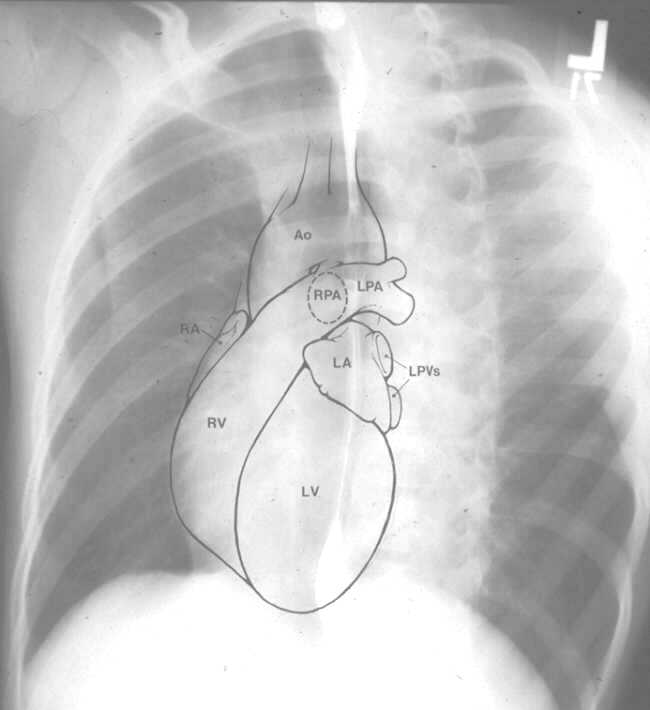
In the left anterior oblique view, the anterior chamber is the right ventricle, and the posterior chamber is the inflow tract of the left ventricle. Increased convexity of the anterior cardiac chamber suggests right ventricular dilation. If the esophagus contains contrast media, a right aortic arch indenting the esophagus can be identified. The main advantage of this view is to evaluate right and left ventricular dilation.
In the lateral view, the right ventricle constitutes the lower half to two-thirds of the anterior cardiac border, and lies behind the sternum. The upper third constitutes the right ventricular outflow tract and the main pulmonary artery. The upper half of the posterior cardiac border consists of the left atrium and the lower half is the left ventricle.